What is the spinal cord cross-section anatomy explained
TL;DR
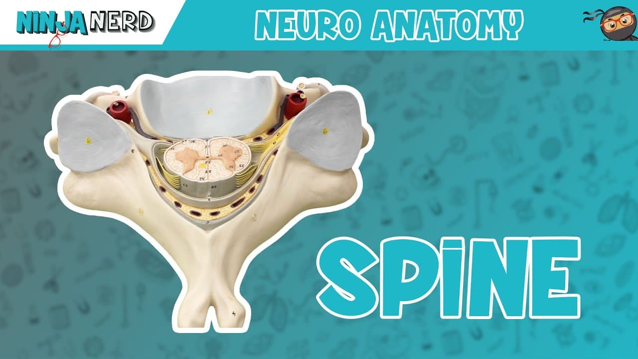
The spinal cord cross-section shows anterior and posterior grey horns, white columns, and a central canal with CSF. It covers the pia mater, arachnoid mater, subarachnoid space, subdural space, and epidural space, plus vertebral arteries and dorsal root ganglia. It explains how somatic nerves exit via the ventral horn and how sensory fibers enter via the dorsal root ganglion.
Transcript
Alright Ninja Nerds. In this video, we're going to talk about the cross-section of the spinal cord. So we're actually looking at c3 through c6. This is actually the body. So this is the anterior side of the cervical vertebrae. These are the superior articulating processes with facets and this is the actual bi-fid spinous process. So this is going t... Read More
Key Insights
- The anterior grey horn houses somatic motor neuron cell bodies that control skeletal muscles.
- The posterior grey horn contains neurons that process peripheral sensory information.
- The central canal contains CSF and is bordered by the gray matter around it.
- The dorsal white column carries proprioception and discriminative touch.
- The lateral white column carries pain and temperature pathways.
- The pia mater clings directly to the spinal cord.
- The subarachnoid space contains cerebrospinal fluid.
- The epidural space lies outside the dura mater and contains fat and vessels.
- More videos from this channel:
- More from this channel: Ninja Nerd
Install to Summarize YouTube Videos and Get Transcripts
Explore YouTube Video Summarizer or Get YouTube Transcript Extractor
Questions & Answers
Q: What is a spinal cord cross-section and what does it show?
A spinal cord cross-section is a horizontal cut through the spinal cord that reveals the organization of gray matter into anterior and posterior horns, and white matter tracts on the outside. It also shows the surrounding meninges, spaces containing CSF, and nearby vascular structures. This view helps link structure to function in motor and sensory pathways.
Q: Where are somatic motor neurons located in the cross-section?
Somatic motor neurons are located in the anterior grey horn of the spinal cord cross-section. These neurons send axons out through the ventral roots to innervate skeletal muscles, forming the basis for voluntary movement. Their position allows integration with sensory input and reflex circuits in the same region.
Q: What roles do the posterior and lateral white columns play?
The posterior white column carries proprioception and discriminative touch, enabling precise sense of limb position and fine tactile differentiation. The lateral white column carries pain and temperature pathways, contributing to the body’s ability to detect and localize nociceptive and thermal stimuli, guiding protective reflexes and conscious perception.
Q: What is the function of the dorsal root ganglion in this model?
The dorsal root ganglion contains unipolar neurons that relay peripheral sensory information into the spinal cord. These first-order neurons transmit signals from receptors to the dorsal horn, where second-order neurons can process and relay to higher brain centers for perception, modulation, and reflex responses.
Q: How are the meninges and spaces arranged around the spinal cord?
From the outside inward, the periosteal dura lines the bone, followed by the epidural space containing fat and vessels. Inside is the arachnoid mater, then the subdural space with a thin barrier, and finally the pia mater clinging to the spinal cord. The subarachnoid space contains cerebrospinal fluid.
Q: What arteries are visible and what do they do?
The vertebral arteries run through transverse foramina and contribute to the posterior part of the Circle of Willis. They supply blood to the posterior circulation of the brain and the spinal cord, ensuring adequate oxygen and nutrient delivery for neural function and metabolism.
Q: How do the ventral and dorsal rami differ in this cross-section?
The ventral ramus contains fibers that exit the anterior grey horn to supply skeletal muscles and skin, contributing to motor and sensory innervation of the ventral body. The dorsal ramus carries fibers that supply the back and the dorsal aspect of the trunk, supporting motor control and sensation in that region.
Q: What key structures define the boundary between CSF spaces and nervous tissue?
The pia mater directly clings to the nervous tissue, forming the innermost layer. The subarachnoid space lies between the pia and arachnoid mater and contains CSF. The subdural space and epidural space lie external to the arachnoid and dura mater, containing potential space fluid and fat, respectively, delineating protection and cushioning for the cord.
Summary & Key Takeaways
-
Summary point 1: The video uses a transverse cross-section to explain how the spinal cord is organized, including anterior and posterior horns and white matter tracts, with attention to orientation and surrounding meninges. It emphasizes the relationship between the cord and the surrounding dura and CSF spaces.
-
Summary point 2: The讲解 covers the layers of the meninges starting from pia mater, then subarachnoid space with CSF, moving to the subdural space, arachnoid mater, and dura mater, and explains the epidural space containing fat and vessels.
-
Summary point 3: The lecture connects anatomy to function by detailing which tracts carry touch, pressure, pain, temperature, proprioception, and discriminative touch, and shows how dorsal root ganglia and ventral horns contribute to sensory and motor pathways.
Read in Other Languages (beta)
Share This Summary 📚
Summarize YouTube Videos and Get Video Transcripts with 1-Click
Try YouTube Summary with ChatGPT & Claude or YouTube Transcript Generator
Explore More Summaries from Ninja Nerd 📚

Summarize YouTube Videos and Get Video Transcripts with 1-Click
Try YouTube Summary with ChatGPT & Claude or YouTube Transcript Generator