How Do Neurons Receive and Transmit Signals?
TL;DR
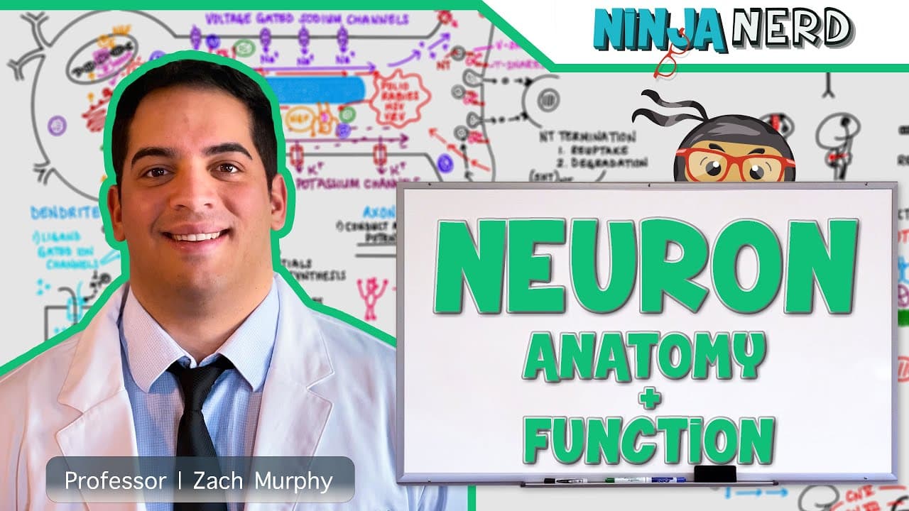
Neurons receive inputs through dendrites and the soma, integrate voltage changes near the axon hillock, and transmit electrical signals along the axon toward its terminals. Ligand-gated channels and G-protein-coupled receptors create graded potentials, while the soma produces neurotransmitters, enzymes, receptors, and other proteins needed for neuronal signaling and cellular activity.
Transcript
all right ninja nerds in this video today we're going to talk about the structure and function of neurons all right guys before you guys get started in this video please hit that like button comment down in the comment section and please subscribe also down in the description box we have links to all our social media platforms for you guys to inter... Read More
Key Insights
- Dendrites are the receptive regions of a neuron, receiving neurotransmitter signals from presynaptic neurons. Their membranes contain ligand-gated ion channels and G-protein-coupled receptors that generate graded changes in membrane voltage and influence whether the neuron becomes more or less likely to fire.
- Ligand-gated ion channels work by opening after a neurotransmitter binds to a receptor pocket. The opened channel permits ions to cross the membrane, changing the electrical state inside the neuron and producing either an excitatory postsynaptic potential or an inhibitory postsynaptic potential.
- An excitatory postsynaptic potential is a depolarizing voltage change that makes the inside of the neuron more positive, or less negative. This graded potential stimulates the neuron toward reaching the threshold required to initiate an action potential.
- An inhibitory postsynaptic potential is a hyperpolarizing voltage change that makes the inside of the neuron more negative than usual and moves it below resting membrane potential. This response opposes excitation and reduces the tendency of the neuron to generate an action potential.
- G-protein-coupled receptors create graded potentials through an indirect signaling pathway. Neurotransmitter binding activates a G protein, second messengers, and protein kinases, which can phosphorylate membrane proteins and alter ion movement to produce either an EPSP or an IPSP.
- The axon hillock is the narrowing region where the soma transitions into the axon. It contains a high concentration of voltage-gated sodium channels, making it the site where action potentials are generated before they move along the axon.
- The cell body is both a receptive region and the main site of neuronal protein synthesis. It produces proteins that can function as neurotransmitters, enzymes, voltage-gated channels, ligand-gated channels, G-protein-coupled receptors, and other components required throughout the cell.
- Nissl bodies are the specialized name for rough endoplasmic reticulum in neurons. They translate mRNA into proteins, which are packaged into vesicles and sent to the Golgi apparatus for modification and additional packaging before use elsewhere in the neuron.
Install to Summarize YouTube Videos and Get Transcripts
Explore YouTube Video Summarizer or Get YouTube Transcript Extractor
Questions & Answers
Q: What are the main structural components of a neuron?
The major structural components are the dendrites, cell body or soma, axon hillock, axon, and axon terminals. Dendrites and the soma receive inputs, the axon hillock is where action potentials are generated, the axon carries those signals, and the bulb-like axon terminals form the final signaling region used for communication with other cells.
Q: How do dendrites receive signals from other neurons?
Dendrites receive signals when presynaptic neurons release neurotransmitters near their membranes. A neurotransmitter can bind directly to a ligand-gated ion channel or activate a G-protein-coupled receptor. Both routes alter membrane proteins and ion movement, producing small graded voltage changes that may excite or inhibit the postsynaptic neuron.
Q: What is the difference between an EPSP and an IPSP?
An EPSP is an excitatory postsynaptic potential that depolarizes the neuron by making its interior more positive, or less negative, and encourages movement toward action-potential threshold. An IPSP is an inhibitory postsynaptic potential that hyperpolarizes the neuron by making its interior more negative than usual, opposing excitation and movement toward threshold.
Q: How do ligand-gated ion channels change neuronal voltage?
A ligand-gated ion channel opens when a neurotransmitter binds to its receptor pocket. Opening removes the functional barrier that normally prevents ion movement across the membrane. Positive ion entry can depolarize the neuron and create an EPSP, while negative ion entry can hyperpolarize it and create an IPSP.
Q: How do G-protein-coupled receptors affect neurons?
A neurotransmitter first binds to the receptive region of a G-protein-coupled receptor. The receptor activates a G protein, which can activate second messengers and protein kinases. Those kinases phosphorylate selected membrane proteins, changing ion flow and generating either an excitatory or inhibitory graded potential through an indirect signaling mechanism.
Q: Why is the axon hillock important for action potentials?
The axon hillock is the funnel-shaped region where the cell body narrows into the axon. It has a high concentration of voltage-gated sodium channels, so it serves as the location where action potentials are generated. Once initiated there, the electrical signal moves away from the soma and down the axon.
Q: What functions does the neuronal cell body perform?
The cell body receives synaptic inputs and participates in the graded potentials that move membrane voltage toward or away from threshold. It also contains the nucleus and protein-synthesis machinery. Through these structures, it makes neurotransmitters, enzymes, receptors, ion channels, and other proteins needed for neuronal signaling and cellular processes.
Q: How are proteins synthesized and packaged inside a neuron?
Protein production begins when a gene in nuclear DNA is transcribed into mRNA. The mRNA leaves the nucleus and reaches the rough endoplasmic reticulum, called Nissl bodies in neurons, where it is translated into protein. The protein is packaged into a vesicle and transferred to the Golgi apparatus for modification and further packaging.
Summary & Key Takeaways
-
A neuron consists of dendrites, a cell body or soma, an axon hillock, an axon, and axon terminals. These specialized regions divide neuronal signaling into reception, integration, electrical transmission, and communication with other neurons. The lecture also identifies myelin and glial support as important parts of understanding neuronal function.
-
Dendrites and the soma receive neurotransmitter signals through ligand-gated ion channels and G-protein-coupled receptors. These interactions produce graded voltage changes called EPSPs or IPSPs. Excitatory inputs depolarize the membrane, while inhibitory inputs hyperpolarize it. The combined changes help determine whether membrane voltage reaches the threshold needed to generate an action potential.
-
The soma contains the nucleus and cellular machinery required for protein synthesis. DNA is transcribed into mRNA, which leaves the nucleus and is translated at rough endoplasmic reticulum, also called Nissl bodies in neurons. Newly formed proteins are packaged and transferred to the Golgi apparatus for modification and further packaging into vesicles.
Read in Other Languages (beta)
Share This Summary 📚
Summarize YouTube Videos and Get Video Transcripts with 1-Click
Try YouTube Summary with ChatGPT & Claude or YouTube Transcript Generator
Explore More Summaries from Ninja Nerd 📚
Summarize YouTube Videos and Get Video Transcripts with 1-Click
Try YouTube Summary with ChatGPT & Claude or YouTube Transcript Generator