How Does Gastrulation Form the Three Germ Layers?
TL;DR
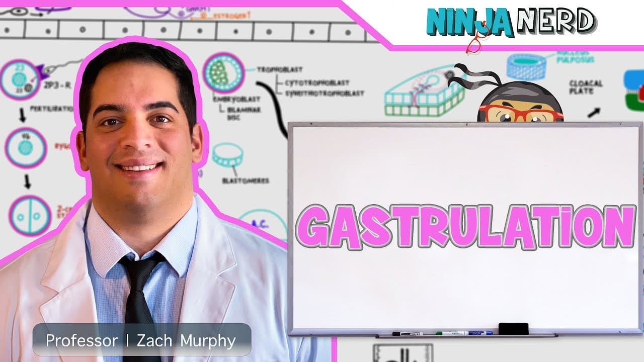
Gastrulation turns the bilaminar embryonic disc into a trilaminar structure made of ectoderm, mesoderm, and endoderm, the three germ layers that build every tissue and organ. It begins with the primitive streak forming on the epiblast surface, after which epiblast cells migrate and invaginate, displacing the hypoblast to create definitive endoderm while remaining epiblast cells become ectoderm.
Transcript
I engineers so we're going to continue talking about the development of the embryo we already finished if you guys haven't already seen it go watch our first video which is the development up until week one right so we talked about fertilization we talked about cleavage and we talked about the formation of the blastocyst right and then how we got t... Read More
Key Insights
- Gastrulation is the process that converts the bilaminar embryo into a trilaminar structure, producing the ectoderm, mesoderm, and endoderm. These three germ layers go on to form every tissue and organ in the body, which is why the event is treated as critical.
- The primitive streak forms on the surface of the epiblast and marks the start of gastrulation. Epiblast cells then migrate toward it and invaginate, and the sequence of that migration determines which germ layer each population of cells ends up in.
- Definitive endoderm is created when migrating epiblast cells displace the hypoblast. Additional layers of migrating cells develop into the intraembryonic mesoderm, and the epiblast cells that remain in place on the surface become the ectoderm.
- The trophoblast, which used to be the outer cell mass, differentiates into the cytotrophoblast and the syncytiotrophoblast. It matters because it becomes part of the chorion and part of the placenta, supplying oxygen and nutrients and removing waste between mother and fetus.
- The cytotrophoblast consists of cells with well-defined margins, each with its nucleus inside its own membrane. It sits at the base of the invading structure and later proliferates into the center of the villus that extends toward the uterine lining.
- The syncytiotrophoblast forms when proliferating cells break down their membranes, spilling cytoplasm and nuclei into a shared pool. The result is a syncytium with no defined cell margins, appearing as finger-like processes extending into the uterine lining.
- Implantation depends on selectins and integrins on the blastocyst that let it hook onto the endometrium. After attaching, it burrows in, and cytotrophoblast cells proliferate through the zona pellucida to begin forming the invading processes.
- Beta hCG, released by the syncytiotrophoblast around day 21 to 24, keeps the corpus luteum producing progesterone. Without it, falling progesterone makes uterine vessels spasm and rupture, the endometrial cells turn necrotic, and the lining is shed along with the embryo.
Install to Summarize YouTube Videos and Get Transcripts
Explore YouTube Video Summarizer or Get YouTube Transcript Extractor
Questions & Answers
Q: What is gastrulation in embryology?
Gastrulation is the developmental event that transforms the bilaminar embryonic disc into a trilaminar structure. It generates the three germ layers, ectoderm, mesoderm, and endoderm, which give rise to every tissue and organ in the body. The process begins with the formation of the primitive streak on the surface of the epiblast, followed by migration and invagination of epiblast cells. Those migrating cells displace the hypoblast to form definitive endoderm, further layers become intraembryonic mesoderm, and the epiblast cells left behind become ectoderm. It occurs as the embryo develops through week two.
Q: How do the three germ layers form during gastrulation?
All three germ layers derive from the epiblast. First, the primitive streak appears on the epiblast surface. Epiblast cells then migrate toward the streak and invaginate. The earliest migrating cells displace the hypoblast and take its place, forming the definitive endoderm. Additional cells that migrate afterward settle between the layers and develop into the intraembryonic mesoderm. The epiblast cells that never migrate remain on the surface and become the ectoderm. This layered ordering is what turns a two-layer disc into a three-layer disc.
Q: What is the difference between the cytotrophoblast and the syncytiotrophoblast?
The trophoblast differentiates into these two layers. The cytotrophoblast is made of cells with well-defined cell margins, each nucleus sitting inside its own intact cell membrane, and it forms the base layer. The syncytiotrophoblast forms when proliferating cells break down their cell membranes so that their cytoplasm and nuclei spill out and fuse into a shared pool, a syncytium with no defined cell boundaries. The syncytiotrophoblast appears as finger-like processes extending outward into the uterine lining, while the cytotrophoblast later proliferates into the center of those processes.
Q: How does the blastocyst implant into the uterine lining?
During cleavage, the developing embryo travels toward the uterine cavity, and the blastocyst pops off into that cavity. It carries specific proteins on its surface, certain selectins and integrins, that allow it to hook up with the endometrium and start implanting. Once attached it burrows into the lining. Cytotrophoblast cells then proliferate and work their way through the zona pellucida, forming finger-like processes. The cells inside those processes disintegrate, spilling cytoplasm and nuclei to form the syncytiotrophoblast, which invades further into the uterine lining.
Q: Why is beta hCG important in early pregnancy?
Beta hCG, human chorionic gonadotropin, is released by the syncytiotrophoblast starting around day 21 to 24. It signals the corpus luteum of the ovary to continue producing progesterone. This matters because progesterone normally drops later in the luteal phase, and when it drops, the vessels within the uterine lining begin spasming and then rupture, the endometrial cells become necrotic, and the lining is shed. If the lining is shed, the implanted embryo goes with it. Beta hCG keeps progesterone elevated so shedding does not occur.
Q: What is the notochord and why does it matter?
The notochord is one of the key midline structures that forms during gastrulation. Its significance is signaling: it plays a pivotal role in signaling for neurulation and for axial patterning of the embryo. Its formation is covered alongside the emergence of the three germ layers, since gastrulation produces both the layered organization of the embryonic disc and the midline landmarks that organize the body axis. Neurulation, the process the notochord helps trigger, follows on from the trilaminar arrangement established by gastrulation.
Q: How does the embryo get oxygen and nutrients from the mother?
The syncytiotrophoblast releases hydrolytic enzymes that work through the uterine lining, where maternal blood vessels are located. It keeps extending until it becomes confluent with those maternal blood vessels. Once that connection is made, the embryo can receive oxygen, nutrients, and hormones, everything it needs to grow, because a link now exists between the embryo and the maternal vascular system through the syncytiotrophoblast and cytotrophoblast. This developing connection becomes the placenta, which also removes waste between the mother and the fetus.
Q: What happens before gastrulation in the first week of development?
The first week covers fertilization, cleavage, and blastocyst formation. LH from the anterior pituitary triggers ovulation, releasing a secondary oocyte frozen in metaphase II, and the sperm meets that oocyte in the ampulla for fertilization. Cleavage then proceeds from the zygote to the 4-cell stage, the 8-cell stage, the 16-cell stage, and finally the blastocyst, all while moving toward the uterine cavity. The outer cell mass becomes the trophoblast and the inner cell mass becomes the embryoblast, which forms the bilaminar disc that gastrulation later converts into three layers.
Summary & Key Takeaways
-
Development up to week one covers fertilization, cleavage, and blastocyst formation. The trophoblast, formerly the outer cell mass, differentiates into the cytotrophoblast and the syncytiotrophoblast, while the inner cell mass becomes the embryoblast, which first forms the bilaminar disc and later the trilaminar disc through gastrulation.
-
LH from the anterior pituitary triggers ovulation, releasing a secondary oocyte arrested in metaphase II. Fertilization occurs in the ampulla, and cleavage proceeds from zygote to 4-cell, 8-cell, and 16-cell stages, then to the blastocyst, all while the developing structure travels toward the uterine cavity for implantation.
-
The blastocyst carries selectins and integrins that let it attach to the endometrium. Cytotrophoblast cells proliferate past the zona pellucida into finger-like processes; their membranes disintegrate, pooling cytoplasm and nuclei into a syncytium. This syncytiotrophoblast releases hydrolytic enzymes and becomes confluent with maternal blood vessels.
-
Around day 21 to 24 the syncytiotrophoblast secretes beta hCG, human chorionic gonadotropin, which signals the corpus luteum to keep producing progesterone. Sustained progesterone stops uterine vessels from spasming and rupturing, preventing necrosis and shedding of the endometrial lining, so the implanted embryo is not shed with it.
-
Gastrulation itself starts with the primitive streak on the epiblast. Migrating and invaginating epiblast cells displace the hypoblast to form definitive endoderm, further layers become intraembryonic mesoderm, and leftover epiblast becomes ectoderm. Midline structures such as the notochord form and signal for neurulation and axial patterning.
Read in Other Languages (beta)
Share This Summary 📚
Summarize YouTube Videos and Get Video Transcripts with 1-Click
Try YouTube Summary with ChatGPT & Claude or YouTube Transcript Generator
Explore More Summaries from Ninja Nerd 📚


Summarize YouTube Videos and Get Video Transcripts with 1-Click
Try YouTube Summary with ChatGPT & Claude or YouTube Transcript Generator