What are the spinal cord segments and nerves?
TL;DR
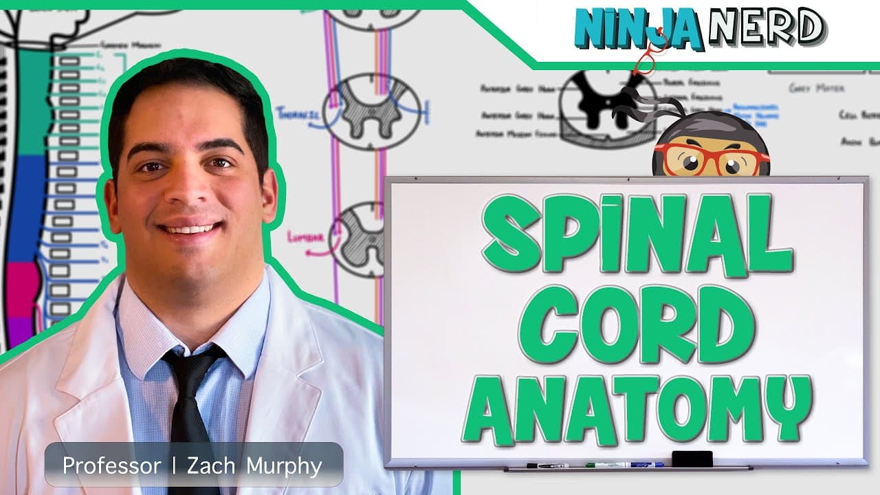
The spinal cord stretches from the foramen magnum to around L1–L2, with cervical, thoracic, lumbar, sacral, and coccygeal segments each giving rise to spinal nerves. There are 31 pairs of spinal nerves, with eight cervical nerves due to exit above their vertebrae, twelve thoracic, five lumbar, five sacral, and one coccygeal. The conus medullaris marks the end, and the cauda equina includes the nerve bundle below it.
Transcript
hi ninja nerds in this video we're going to talk about the gross anatomy of the spinal cord and the spinal nerves let's go and get started all right ninja so what we have to do first is we have to talk about the spinal cord right and its associated spinal nerves now before we do that i just want to make a little quick division over here you guys re... Read More
Key Insights
- The spinal cord is organized into cervical, thoracic, lumbar, sacral, and coccygeal segments.
- The spinal nerves arise from these segments and exit the vertebral column between adjacent vertebrae.
- The first cervical nerve exits above its corresponding vertebra, while subsequent nerves exit below the vertebrae.
- The conus medullaris marks the tapered end of the spinal cord, around L1–L2.
- The cauda equina is a bundle of spinal nerves below the conus medullaris, resembling a horse tail.
- There are 31 pairs of spinal nerves in total, distributed as 8 cervical, 12 thoracic, 5 lumbar, 5 sacral, and 1 coccygeal.
- Spinal nerves carry both sensory and motor information, with dorsal root ganglia acting as sensory relays.
- Embryological fusion creates a single sacrum from five vertebrae, evidenced by transverse ridges.
- More videos from this channel:
- More from this channel: Ninja Nerd
Install to Summarize YouTube Videos and Get Transcripts
Explore YouTube Video Summarizer or Get YouTube Transcript Extractor
Questions & Answers
Q: How many spinal nerve pairs are there and how are they distributed across segments?
There are 31 pairs of spinal nerves in total. Eight pairs arise in the cervical region, twelve in the thoracic region, five in the lumbar region, five in the sacral region, and one in the coccygeal region. This distribution matches the segmental organization of the spinal cord, with each segment giving rise to its corresponding spinal nerves that exit between adjacent vertebrae. The exception is the cervical region where the first eight nerves exit above or near their vertebrae rather than strictly below.
Q: Where does the spinal cord begin and end, and what anatomical landmarks define this range?
The spinal cord begins at the foramen magnum, where the brainstem connects to the cord, and ends at the conus medullaris around the L1–L2 level. This range defines the primary central nervous system structure that houses the spinal cord and its segmental organization. The end of the cord gives way to the cauda equina, a bundle of nerves that continues within the vertebral canal.
Q: What is the conus medullaris and where is it located?
The conus medullaris is the tapered end of the spinal cord, typically located around the L1–L2 vertebral level. It marks the termination of the spinal cord itself, after which only nerve roots continue within the vertebral canal. Understanding its location helps explain why the cauda equina forms below it and how nerve roots descend to exit at lower levels.
Q: What is the cauda equina and why is it named that?
The cauda equina is a bundle of spinal nerves that extends from the end of the spinal cord to the coccygeal level, roughly from L2 to coccygeal 1. It is called the horse tail because the collection of nerve roots resembles a tail of a horse when viewed caudally. This structure allows nerves to reach the lower spinal levels despite the shorter spinal cord.
Q: How many cervical vertebrae and why are there eight cervical spinal nerves?
There are eight cervical spinal nerve pairs because the first cervical nerve exits above its corresponding vertebra, and the eighth cervical nerve exits below the second cervical vertebra yet above the first thoracic vertebra. From C2 downward, each nerve exits below its corresponding vertebra. This exit pattern creates eight cervical nerves despite seven cervical vertebrae. The arrangement differs for thoracic and lower segments where nerves exit below their vertebrae.
Q: What are the major external features used to describe the spinal cord’s anatomy?
Major external features include the anterior median fissure, the posterior median sulcus, and the gray matter horns surrounded by white matter columns. The dorsal and ventral roots connect to these regions, with dorsal root ganglia associated with sensory input. These features help map motor and sensory pathways to specific spinal segments, clarifying functional organization within the spinal cord.
Q: How are the spinal nerves classified in relation to the peripheral nervous system?
Spinal nerves are part of the peripheral nervous system and consist of sensory and motor components, which can be somatic or autonomic in function. These nerves emerge from the spinal cord, carry signals to and from the body, and are divided into dorsal (sensory) and ventral (motor) roots. Their organization allows integration of reflexes, voluntary actions, and autonomic control.
Q: What embryological fact explains the sacrum being a single bone formed from multiple vertebrae?
Embryologically, the sacrum forms from five initially separate vertebrae that fuse into a single bone. The fusion process leaves transverse ridges visible on the bone indicating the original individual elements. This developmental background explains why there are five sacral nerve pairs even though the sacrum is a single bone in the adult.
Summary & Key Takeaways
-
The spinal cord is part of the central nervous system and is connected to the peripheral nervous system via spinal nerves.
-
It starts at the foramen magnum and ends at the conus medullaris around L1–L2, with distinct cervical to coccygeal segments.
-
There are 31 pairs of spinal nerves total, with eight cervical nerves explained by exit above vertebrae.
-
The cauda equina is a bundle of nerves below the end of the spinal cord resembling a horse tail.
-
Embryologically the sacrum is one bone but formed from five fused vertebrae, noted by transverse ridges.
-
The spinal nerves exit between vertebrae and include dorsal root ganglia and branching patterns.
-
Dorsal and ventral roots carry sensory and motor information, linking spinal nerves to body functions.
-
Enlargements of the spinal cord relate to limb innervation and segmental organization.
Read in Other Languages (beta)
Share This Summary 📚
Summarize YouTube Videos and Get Video Transcripts with 1-Click
Try YouTube Summary with ChatGPT & Claude or YouTube Transcript Generator
Explore More Summaries from Ninja Nerd 📚






Summarize YouTube Videos and Get Video Transcripts with 1-Click
Try YouTube Summary with ChatGPT & Claude or YouTube Transcript Generator