How Does the Embryonic GI Tract Develop?
TL;DR
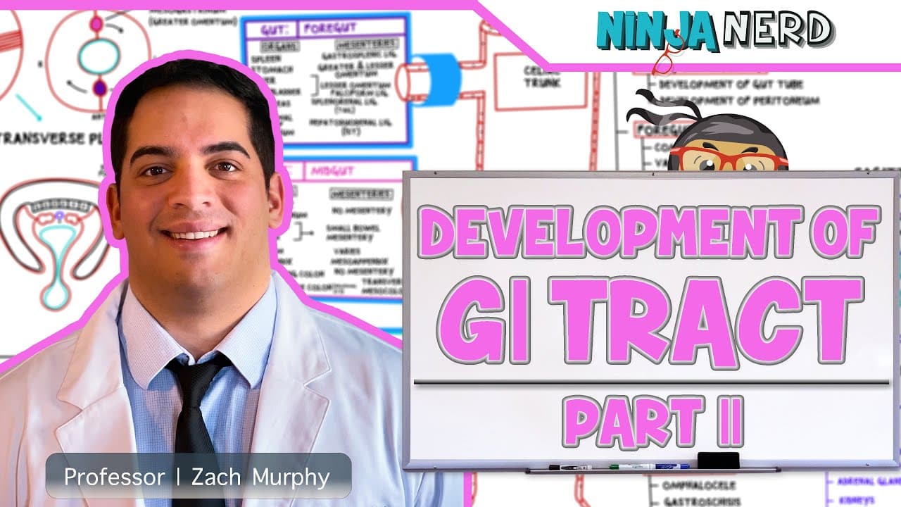
The embryonic gut divides into the foregut, midgut, and hindgut, with each region producing distinct structures and receiving a characteristic arterial supply. Mesenteries suspend organs while carrying blood vessels, veins, nerves, and lymphatics, and developmental rotation rearranges the stomach, liver, spleen, intestines, and peritoneal spaces into their final relationships.
Transcript
iein engineers in this video we're gonna talk about the development of the GI tract this is part two if you haven't go and see part one alright because in part one we talked about the embryonic folding we talked about the intraperitoneal retroperitoneal and pathologies right in this video we're going to be specifically focusing on the foregut the m... Read More
Key Insights
- A mesentery is a double-layered serous membrane composed of simple squamous epithelial tissue with underlying areolar connective tissue, described as mesothelium. It connects the visceral peritoneum surrounding an organ to the parietal peritoneum lining the abdominal wall.
- Mesenteries are pathways for an organ's blood vessels, draining veins, nerve fibers, and lymphatic vessels. These structures travel between the abdominal wall and the organ through the potential space created within the double-layered membrane.
- The foregut is distinctive because it possesses two mesenteries, the ventral mesogastrium on the anterior side and the dorsal mesogastrium on the posterior side. The liver develops within the ventral mesogastrium, while the spleen develops within the dorsal mesogastrium.
- Foregut rotation is a clockwise movement involving the stomach and proximal portion of the duodenum around a central axis. This rotation shifts the developing liver from its anterior position toward the right side and positions the developing spleen on the left side.
- The ventral mesogastrium forms the lesser omentum and contributes to peritoneal attachments including the hepatoduodenal, hepatogastric, and falciform ligaments. These structures help maintain relationships among intraperitoneal organs derived from the foregut region.
- The dorsal mesogastrium forms the greater omentum and contributes to attachments such as the gastrocolic, gastrosplenic, and splenorenal ligaments. These named ligaments are peritoneal coverings rather than the bone-to-bone ligaments found in the musculoskeletal system.
- The greater omentum is an apron-like structure that hangs from the stomach's greater curvature, descends over the intestines, and doubles back. During development, it fuses with portions of the transverse colon's mesenteric attachments.
- The gut tube has three divisions called the foregut, midgut, and hindgut, supplied respectively by the celiac trunk, superior mesenteric artery, and inferior mesenteric artery. The midgut herniates, rotates 270 degrees counterclockwise, and returns to the abdominal cavity.
Install to Summarize YouTube Videos and Get Transcripts
Explore YouTube Video Summarizer or Get YouTube Transcript Extractor
Questions & Answers
Q: What is a mesentery in gastrointestinal embryology?
A mesentery is a double-layered serous membrane that connects the visceral peritoneum covering an organ with the parietal peritoneum lining the abdominal wall. Its layers contain simple squamous epithelial tissue and underlying areolar connective tissue. The space within it provides a route for blood vessels, draining veins, splanchnic nerve fibers, and lymphatic vessels traveling to or from an organ.
Q: Why does the foregut have two mesenteries?
The foregut is distinctive among the three gut divisions because it has both an anterior ventral mesogastrium and a posterior dorsal mesogastrium. The liver develops within the ventral mesogastrium, while the spleen develops within the dorsal mesogastrium. As foregut structures rotate, these mesenteries are rearranged and produce the lesser omentum, greater omentum, and several associated peritoneal attachments.
Q: How does foregut rotation position the liver and spleen?
The stomach and proximal portion of the duodenum rotate clockwise around a central axis during foregut development. This movement carries the liver from its earlier anterior position toward the right side of the body. At the same time, the spleen, which develops within the dorsal mesogastrium, becomes positioned on the left side.
Q: What develops from the ventral mesogastrium?
The ventral mesogastrium produces the lesser omentum and contributes to several peritoneal attachments, including the hepatoduodenal ligament, hepatogastric ligament, and falciform ligament. The liver develops within this ventral mesentery before rotation changes its position. These structures help preserve connections among foregut organs and support their intraperitoneal relationships.
Q: What develops from the dorsal mesogastrium?
The dorsal mesogastrium contributes to the greater omentum and associated peritoneal attachments, including the gastrocolic, gastrosplenic, and splenorenal ligaments. The spleen develops within this posterior mesentery. As the stomach rotates and the mesentery lengthens, the greater omentum descends from the greater curvature, doubles back, and later fuses near the transverse mesocolon.
Q: What are the greater and lesser omenta?
The lesser omentum connects the stomach's lesser curvature and proximal duodenum with the liver and includes the hepatogastric and hepatoduodenal ligaments. The greater omentum extends from the stomach's greater curvature, hangs over the intestines like an apron, and doubles back. Its returning layers fuse with tissues associated with the transverse colon and transverse mesocolon.
Q: What are the lesser sac and epiploic foramen?
The lesser sac is a peritoneal space located behind the stomach. It is related to the arrangement created as the greater omentum and surrounding mesenteries develop. The epiploic foramen, also called the epiploic foramen of Winslow, is an opening near the end of the hepatoduodenal ligament that provides access into the lesser sac.
Q: How do the foregut, midgut, and hindgut differ?
The embryonic gut tube is divided into the foregut, midgut, and hindgut, with each division having distinct components and vascular organization. Their arterial supplies are the celiac trunk, superior mesenteric artery, and inferior mesenteric artery, respectively. The midgut also undergoes herniation, rotates 270 degrees counterclockwise, and then returns to the abdominal cavity during development.
Summary & Key Takeaways
-
The foregut is unusual because it has both ventral and dorsal mesenteries, called the ventral mesogastrium and dorsal mesogastrium. These double-layered serous membranes connect visceral and parietal peritoneum while providing routes through which blood vessels, veins, nerve fibers, and lymphatic vessels can reach developing abdominal organs.
-
As the stomach and proximal duodenum rotate clockwise around their central axis, the developing liver moves toward the right and the spleen moves toward the left. The ventral mesogastrium forms the lesser omentum and associated peritoneal ligaments, while the dorsal mesogastrium contributes to the greater omentum and its related attachments.
-
The greater omentum descends from the stomach's greater curvature, doubles back, and fuses with tissues around the transverse mesocolon. Behind the stomach, it helps define the lesser sac. The lesser sac can be entered through the epiploic foramen near the end of the hepatoduodenal ligament.
-
Key Insights
Read in Other Languages (beta)
Share This Summary 📚
Summarize YouTube Videos and Get Video Transcripts with 1-Click
Try YouTube Summary with ChatGPT & Claude or YouTube Transcript Generator
Explore More Summaries from Ninja Nerd 📚


Summarize YouTube Videos and Get Video Transcripts with 1-Click
Try YouTube Summary with ChatGPT & Claude or YouTube Transcript Generator