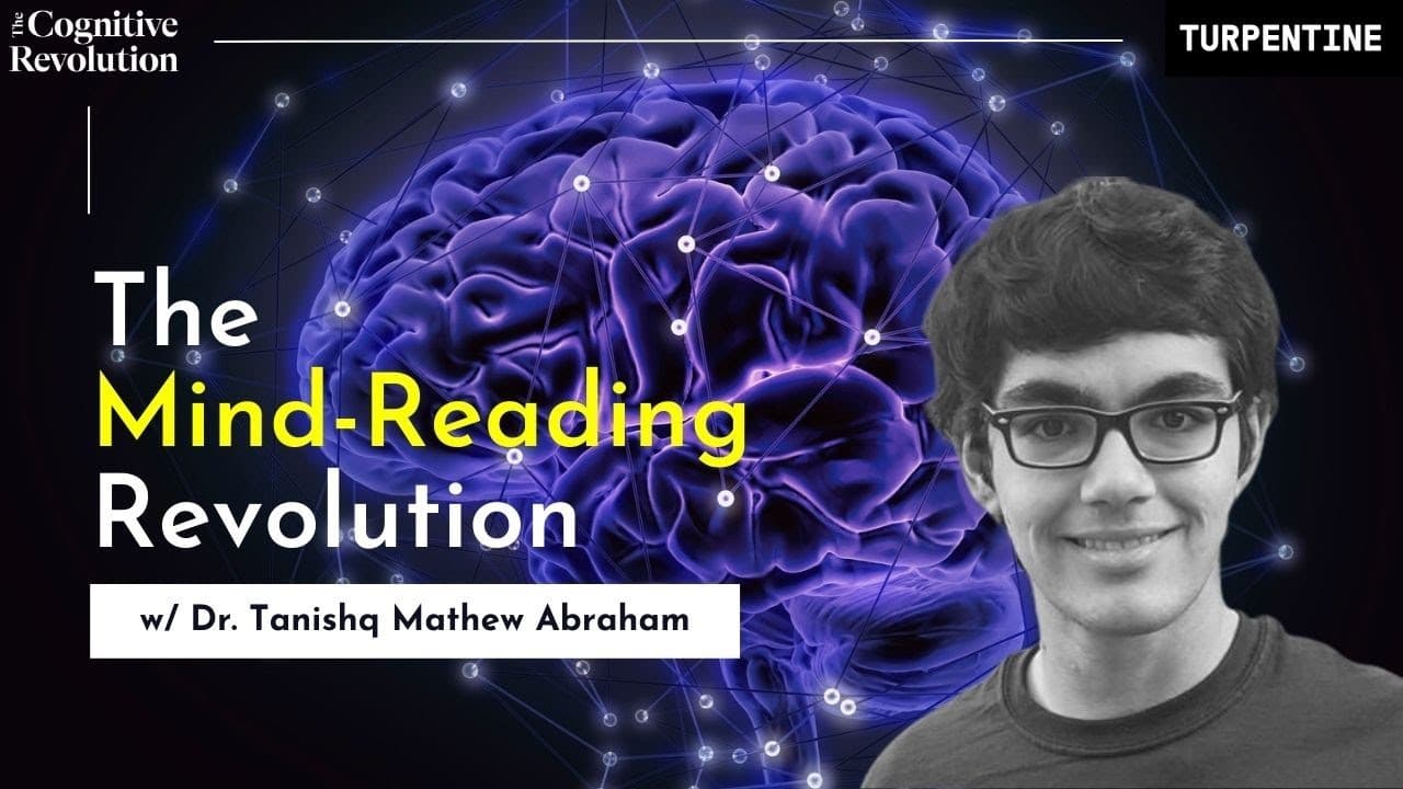
How Does AI Reconstruct Images from fMRI? The Cognitive Revolution with Dr. Tanishq Mathew Abraham (Part 1 of 2)
TL;DR
AI reconstructs what a person viewed by mapping fMRI brain activity into the representation spaces of pre-trained CLIP and Stable Diffusion models. The approach combines semantic and low-level image representations, uses roughly 15,000 voxel data points, and trains a separate model for each person. Read on to understand how this works with limited data and why it could advance neuroscience.
Transcript
I think this idea of of mapping one latent space to another is a very powerful idea I think it's always best to try to take advantage of that as much as possible and the real I guess Innovation these days is to be able to use these multimodal spaces as well um and being able to map you know different things to these to these multimodal spaces that'... Read More
Key Insights
- AI can reconstruct visual perceptions from fMRI data, mapping brain activity to images.
- The key is leveraging pre-trained models like CLIP and stable diffusion for image generation.
- Mapping brain data to a shared representation space allows for semantic understanding.
- fMRI scans provide 15,000 voxel data points, representing brain activity during image viewing.
- Separate models are trained for each individual due to unique brain activity patterns.
- Low data environments can still yield breakthrough results with thoughtfully designed architectures.
- Potential applications include understanding brain function and aiding neurological diagnostics.
- The approach highlights the power of mapping between high-dimensional spaces in AI research.
Install to Summarize YouTube Videos and Get Transcripts
Explore YouTube Video Summarizer or Get YouTube Transcript Extractor
Questions & Answers
Q: How does AI reconstruct images from fMRI brain scans?
The system takes fMRI data recorded while a person views an image and maps that brain activity into pre-trained model spaces. CLIP and Stable Diffusion then help reconstruct an image that reflects the original visual stimulus.
Q: What is “Reconstructing the Mind’s Eye”?
“Reconstructing the Mind’s Eye” is the first paper discussed with Dr. Tanishq Mathew Abraham on The Cognitive Revolution. Its full topic is fMRI-to-image reconstruction using contrastive learning and diffusion priors.
Q: What roles do CLIP and Stable Diffusion play in the reconstruction process?
CLIP provides a pre-trained semantic representation space to which the fMRI data can be mapped. Stable Diffusion uses the resulting guidance to generate a visual reconstruction of what the person viewed.
Q: What information does an fMRI scan provide to the model?
The fMRI scan supplies about 15,000 voxel data points representing brain activity while the subject views an image. The model translates that activity into representations that can guide image reconstruction.
Q: Why are separate AI models trained for each person?
Separate models are used because measured brain-activity patterns differ between individuals. Personalizing the mapping helps connect each person’s fMRI voxel data with the corresponding visual stimuli.
Q: How can the method work with relatively small datasets and modest compute budgets?
The researchers leverage pre-trained foundation models rather than learning every representation from scratch. Thoughtfully designed, problem-specific architectures map limited fMRI data into the rich spaces already provided by CLIP and Stable Diffusion.
Q: Why is mapping between latent or multimodal spaces important?
Mapping lets the system connect different kinds of information, including brain activity and visual representations. Dr. Abraham describes mapping one latent space to another, and mapping different inputs into multimodal spaces, as a powerful approach.
Q: How could fMRI-to-image reconstruction advance neuroscience?
It offers a non-invasive way to decode brain activity and study how people process visual information. The existing page also identifies possible applications in understanding cognition, neurological diagnostics, and personalized brain-computer interfaces.
Summary & Key Takeaways
-
AI technology has advanced to a point where it can reconstruct images from brain activity data, specifically using fMRI scans. This is achieved by mapping the brain's voxel data onto pre-trained model spaces, allowing for image generation that reflects the original visual stimuli. Such work demonstrates the potential for AI to aid in neuroscience research and diagnostics.
-
The process involves using a combination of semantic and low-level image representations to guide the reconstruction of images from brain data. By training separate models for each individual, researchers can account for unique brain activity patterns, paving the way for personalized brain-computer interfaces.
-
This research underscores the importance of pre-trained models and high-dimensional space mapping in AI. Despite the challenges of low data environments, the innovative use of existing models enables significant progress, opening new doors for understanding human cognition and potential clinical applications.
Read in Other Languages (beta)
Share This Summary 📚
Summarize YouTube Videos and Get Video Transcripts with 1-Click
Try YouTube Summary with ChatGPT & Claude or YouTube Transcript Generator
Explore More Summaries from Cognitive Revolution "How AI Changes Everything" 📚





Summarize YouTube Videos and Get Video Transcripts with 1-Click
Try YouTube Summary with ChatGPT & Claude or YouTube Transcript Generator