How Does the Female Reproductive System Work? | Anatomy Model
TL;DR
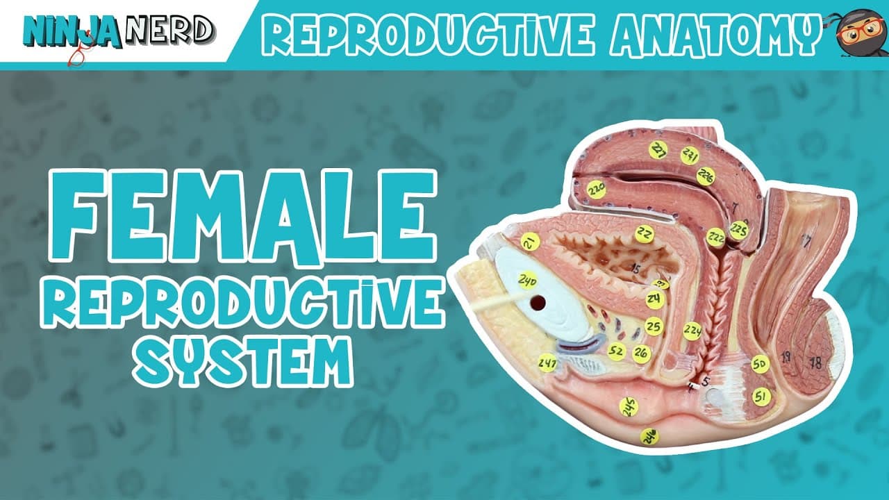
The female reproductive system works through connected organs that produce and transport oocytes, support fertilization, and anchor the uterus. The ovaries contain maturing follicles, fimbriae guide a released secondary oocyte into the uterine tube, and fertilization occurs in its ampulla. The anatomy model also identifies four uterine regions and several supporting ligaments, making the detailed structure-by-structure explanation worth reading.
Transcript
all right engineers in this video we're going to talk about the female reproductive system all right so if you guys look here we're going to look at this from the anterior view this right here this fatty rounded region right here is called the mons pubis okay and it's usually got going to be covered with hair might not be within all people right yo... Read More
Key Insights
- The uterus is organized into the fundus, body, isthmus, and cervix. The fundus is the rounded superior region, the body forms the main portion, and the isthmus and cervix occupy the lower part, although some divisions are difficult to see clearly on this model.
- The ovary is the site where ovarian follicles mature and help produce the female oocyte. It connects to the uterus through the ovarian ligament, while surrounding ligament structures provide additional lateral support for the reproductive organs.
- The broad ligament is described as a large supporting structure made up of the mesovarium and suspensory ligaments. It helps anchor the reproductive structures laterally, in contrast with the anterior support of the round ligament and posterior support of the uterosacral ligament.
- The uterosacral ligament anchors the uterus toward the sacral region posteriorly. The lecture also calls it the sacral uterine ligament, presenting the two names as interchangeable descriptions of the same supporting connection.
- The fimbriae are fingerlike projections that stiffen and sweep across the ovarian surface near ovulation. Their role is to help capture the released secondary oocyte and move it toward the opening of the uterine tube.
- The ampulla is a dilated region of the uterine tube where fertilization occurs. It lies behind the infundibulum, the stalklike portion from which the fimbriae extend, while the tube continues toward the uterus through its isthmus.
- The external genital structures include the hair-bearing labia majora, the hairless labia minora, the vestibule, and the clitoris. The clitoris contains erectile tissue, numerous blood vessels, and many nerve fibers, allowing it to become engorged during tactile or erotic stimulation.
- The structure identified at 6:05 as a Bartholin gland is actually the bulbus vestibuli, according to the description's correction. The bulbus vestibuli belongs to erectile tissue and the bulboclitoral organ, while the greater vestibular glands lie more dorsally and perform another function.
Install to Summarize YouTube Videos and Get Transcripts
Explore YouTube Video Summarizer or Get YouTube Transcript Extractor
Questions & Answers
Q: How does the female reproductive system work?
The ovaries contain follicles that mature and help produce female oocytes. Near ovulation, fimbriae sweep across the ovary and guide a released secondary oocyte toward the uterine tube, where fertilization occurs in the ampulla. The uterus and its supporting ligaments form another major part of this reproductive anatomy.
Q: What are the main parts of the uterus?
The uterus is divided into the fundus, body, isthmus, and cervix. The fundus is its rounded superior region, while the body forms the main portion. The isthmus and cervix occupy the lower part, although they are difficult to distinguish on the first anatomy model.
Q: Which ligaments support the uterus and ovaries?
The ovarian ligament connects the ovary directly to the uterus. The round ligament anchors the uterus anteriorly, the uterosacral ligament anchors it posteriorly toward the sacral region, and the broad ligament provides lateral support. The broad ligament is described as including the mesovarium and suspensory ligaments.
Q: How do the fimbriae help move an oocyte?
Near ovulation, the ovary releases a secondary oocyte in metaphase II. The fingerlike fimbriae stiffen and sweep across the ovarian surface, helping capture the released cell and move it toward the uterine tube. They extend from the stalklike infundibulum.
Q: Where does fertilization occur in the uterine tube?
Fertilization occurs in the ampulla of the uterine tube. The ampulla is a dilated region located behind the infundibulum, the stalklike portion from which the fimbriae extend.
Q: What is the difference between the labia majora and labia minora?
The labia majora are the outer skin folds and are described as hair-bearing. The labia minora are the inner, hairless folds associated with the vestibule. The greater vestibular glands secrete mucus into this area.
Q: What is the clitoris made of, and how does it respond to stimulation?
The clitoris contains erectile tissue, numerous blood vessels, and many nerve fibers. During tactile or erotic stimulation, the tissue fills with blood and becomes engorged. A small skin fold called the prepuce surrounds it.
Q: What anatomical labeling error is corrected in the description?
At 6:05, structure number 248 is incorrectly identified as a Bartholin gland. It is actually the bulbus vestibuli, erectile tissue belonging to the bulboclitoral organ. The Bartholin glands, also called greater vestibular glands, are located more dorsally and have a different function.
Summary & Key Takeaways
-
The model presents the female reproductive system from anterior, lateral, and internal views. Major structures include the mons pubis, ovaries, uterine tubes, uterus, cervix, labia, vestibule, clitoris, urethra, and bladder. The lecture repeatedly connects each visible anatomical feature with its location, structural relationships, and function.
-
The uterus is divided into the fundus, body, isthmus, and cervix. Its position is supported by multiple ligaments: the round ligament anchors it anteriorly, the uterosacral ligament anchors it posteriorly, and the broad ligament supports it laterally. The ovarian ligament connects each ovary directly to the uterus.
-
The uterine tube includes the fimbriae, infundibulum, ampulla, and isthmus. Fimbriae help sweep a released secondary oocyte toward the tube, while the ampulla is identified as the site of fertilization. The description also corrects the model demonstration by distinguishing the bulbus vestibuli from the more dorsally located Bartholin glands.
Read in Other Languages (beta)
Share This Summary 📚
Summarize YouTube Videos and Get Video Transcripts with 1-Click
Try YouTube Summary with ChatGPT & Claude or YouTube Transcript Generator
Explore More Summaries from Ninja Nerd 📚



Summarize YouTube Videos and Get Video Transcripts with 1-Click
Try YouTube Summary with ChatGPT & Claude or YouTube Transcript Generator