How to Distinguish UMN and LMN Lesions
TL;DR
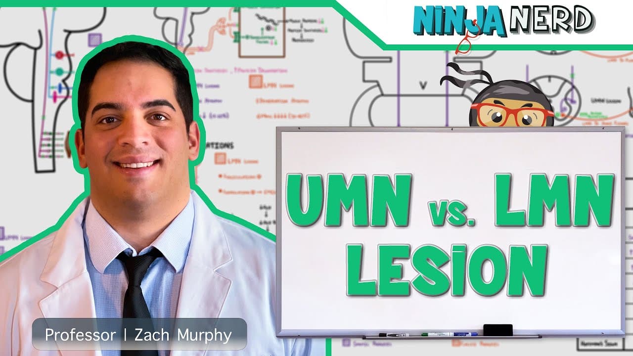
Upper motor neuron lesions produce spasticity, hyperreflexia, clonus, and pathologic reflexes such as the Babinski sign, while lower motor neuron lesions cause flaccid paralysis, hyporeflexia, fasciculations, and muscle atrophy. Distinguishing them requires locating damage along the corticospinal or corticobulbar pathways versus the anterior horn cells, motor cranial nerve nuclei, peripheral axons, or axon terminals.
Transcript
all right engineers so in this video we are going to talk about upper motor neuron versus lower motor neuron lesions let's go and get started all right so when we talk about upper motor neuron versus lower motor neural lesions we first have to understand what in the heck is an upper motor neuron and what is a lower motor neuron now to make this ver... Read More
Key Insights
- Upper motor neurons originate in motor areas of the cerebral cortex and descend through the corona radiata and internal capsule. Their axons continue through the brainstem within corticospinal or corticobulbar pathways before communicating with lower motor neurons.
- The corticospinal tract connects the cerebral cortex with lower motor neuron cell bodies in the anterior gray horn of the spinal cord. Those lower motor neurons then send axons to skeletal muscles of the upper limbs, lower limbs, or trunk.
- The corticobulbar tract connects cortical motor regions with motor cranial nerve nuclei in the pons and medulla. Its destinations include motor nuclei associated with cranial nerves five, seven, nine, ten, the cranial component of eleven, and twelve.
- Lower motor neurons comprise the motor neuron cell body, its axon, and its axon terminal bulb. Damage to a motor cranial nerve nucleus, an anterior horn cell, a peripheral motor axon, or its terminal therefore qualifies as a lower motor neuron lesion.
- Upper motor neuron lesions are marked by spasticity, hyperreflexia, clonus, and pathologic reflexes such as the Babinski sign. Increased muscle tone occurs because damage to the upper pathway removes inhibitory modulation from lower motor circuitry.
- Lower motor neuron lesions are marked by flaccid paralysis, hyporeflexia, fasciculations, and muscle atrophy. These findings result from direct damage to the motor unit anywhere from its cell body through its axon and terminal connection with skeletal muscle.
- Upper motor neuron lesion causes include hemorrhagic or ischemic stroke, multiple sclerosis, Friedreich's ataxia, vitamin B12 deficiency, and amyotrophic lateral sclerosis. The lecture identifies stroke as a principal cause and groups several other examples around demyelination or motor neuron disease.
- Lower motor neuron lesion causes include poliomyelitis, West Nile virus, spinal muscular atrophy, cauda equina compression, diabetic neuropathy, and botulism. These conditions can affect anterior gray horn cells, nerve roots, peripheral axons, or acetylcholine release at axon terminals.
Install to Summarize YouTube Videos and Get Transcripts
Explore YouTube Video Summarizer or Get YouTube Transcript Extractor
Questions & Answers
Q: What is the difference between an upper and lower motor neuron?
An upper motor neuron begins in a motor region of the cerebral cortex and descends through the corticospinal or corticobulbar pathway to communicate with a lower motor neuron. A lower motor neuron consists of a motor cell body in the anterior gray horn or a motor cranial nerve nucleus, plus its axon and terminal extending to skeletal muscle.
Q: How can upper and lower motor neuron lesions be distinguished clinically?
Upper motor neuron lesions are identified by spasticity, hyperreflexia, clonus, and pathologic reflexes such as the Babinski sign. Lower motor neuron lesions instead produce flaccid paralysis, hyporeflexia, fasciculations, and muscle atrophy. These contrasting examination findings help localize dysfunction to descending motor pathways or to the lower motor neuron and its peripheral connection with muscle.
Q: Where can an upper motor neuron lesion occur?
An upper motor neuron lesion can occur anywhere along the descending pathway that begins in the motor areas of the cerebral cortex. This route passes through the corona radiata, internal capsule, midbrain, pons, and medulla, then continues toward the spinal cord through the corticospinal tract or toward motor cranial nerve nuclei through the corticobulbar tract.
Q: Where can a lower motor neuron lesion occur?
A lower motor neuron lesion can affect the cell body, axon, nerve, or axon terminal of the neuron that directly supplies skeletal muscle. Relevant cell bodies are located in the anterior gray horn of the spinal cord or in specific motor cranial nerve nuclei. Damage at any point from those structures to the muscle is classified as a lower motor neuron lesion.
Q: What are common causes of upper motor neuron lesions?
The lecture presents stroke, including hemorrhagic and ischemic cerebrovascular accidents, as a major cause of upper motor neuron damage. It also identifies multiple sclerosis, Friedreich's ataxia, and vitamin B12 deficiency as conditions that can demyelinate upper motor neuron axons. Amyotrophic lateral sclerosis can also damage upper motor neuron structures as part of motor neuron disease.
Q: What are common causes of lower motor neuron lesions?
Lower motor neuron lesions can arise from poliomyelitis, West Nile virus, or autosomal recessive spinal muscular atrophy affecting anterior gray horn cells. Cauda equina syndrome, herniated discs, or spinal stenosis can compress nerve roots or axons. Diabetic neuropathy can damage peripheral axons, while botulism inhibits acetylcholine release at the axon terminal.
Q: Which cranial nerve nuclei receive corticobulbar input?
The described corticobulbar destinations include the motor nucleus associated with cranial nerve five for muscles of mastication and cranial nerve seven for facial expression. The nucleus ambiguus contributes to cranial nerves nine, ten, and the cranial component of eleven for the pharynx, larynx, soft palate, and uvula. Cranial nerve twelve supplies tongue muscles.
Q: Why do UMN and LMN lesions cause different muscle tone findings?
Upper motor neuron lesions increase tone and produce spasticity because descending damage removes inhibitory modulation from lower motor circuitry. Lower motor neuron lesions decrease tone and cause flaccid paralysis because they directly damage the motor unit connecting its cell body to skeletal muscle. The same distinction helps explain hyperreflexia in upper lesions and hyporeflexia in lower lesions.
Summary & Key Takeaways
-
Upper motor neurons begin in motor regions of the cerebral cortex. Their axons descend through the corona radiata and internal capsule, then continue through the brainstem as corticospinal or corticobulbar pathways. The corticospinal tract reaches anterior gray horn cells, while the corticobulbar tract reaches selected motor cranial nerve nuclei.
-
Lower motor neurons include anterior gray horn cells and motor cranial nerve nuclei, along with their axons and terminal bulbs extending to skeletal muscles. The relevant cranial nerve pathways control mastication, facial expression, the pharynx, larynx, soft palate, uvula, and tongue, supporting movement, swallowing, speech, and articulation.
-
Upper motor neuron lesions are associated with spasticity, hyperreflexia, clonus, and pathologic reflexes, whereas lower motor neuron lesions cause flaccid paralysis, hyporeflexia, fasciculations, and muscle atrophy. Common discussed causes include stroke, demyelinating conditions, ALS, viral damage, spinal muscular atrophy, nerve compression, diabetic neuropathy, and botulism.
Read in Other Languages (beta)
Share This Summary 📚
Summarize YouTube Videos and Get Video Transcripts with 1-Click
Try YouTube Summary with ChatGPT & Claude or YouTube Transcript Generator
Explore More Summaries from Ninja Nerd 📚




Summarize YouTube Videos and Get Video Transcripts with 1-Click
Try YouTube Summary with ChatGPT & Claude or YouTube Transcript Generator