How Does Lung and Pleural Anatomy Work?
TL;DR
Air travels through progressively smaller airways until it reaches the alveoli, where oxygen enters pulmonary vessels and carbon dioxide leaves them. The right lung has three lobes, while the left has two, and each contains surgically useful bronchopulmonary segments. Pleural layers, recesses, blood vessels, lymphatic pathways, and autonomic innervation complete the anatomical framework.
Transcript
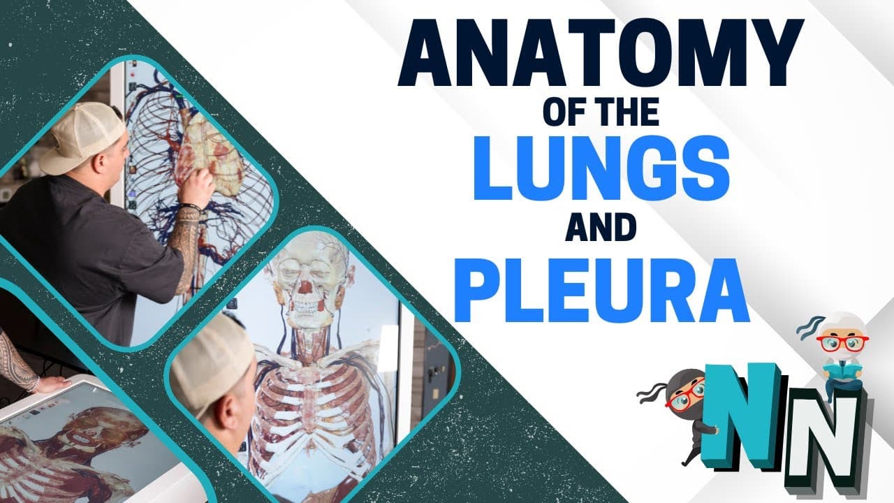
What's up, ninja nerds? In this video today, we're going to be talking about the lungs and the plura. And again, we're going to be utilizing our trusty friend here, the anatomage table to take a look at three-dimensional images. But also, again, remember, we're taking this into a consideration that this is a hybrid. We're gonna have a lot of our di... Read More
Key Insights
- The conducting zone transports air without performing gas exchange. Its pathway includes the trachea, primary bronchi, lobar bronchi, segmental bronchi, bronchioles, and terminal bronchioles before air reaches the respiratory portion of the lung.
- The respiratory zone begins at the respiratory bronchioles and ends with the alveoli. Respiratory bronchioles feed alveolar ducts, which lead into alveolar sacs composed of individual alveoli surrounded by a pulmonary capillary network.
- The right lung has three lobes, while the left lung has two. This arrangement corresponds to three right lobar bronchi, named superior, middle, and inferior, and two left lobar bronchi, named superior and inferior.
- Bronchopulmonary segments are subdivisions supplied through segmental bronchi and separated from neighboring segments by connective tissue septa. This separation makes it possible to remove a diseased segment while preserving other segments within the same lobe.
- Alveoli are the structural and functional units of the lungs. Their thin simple squamous epithelial structure and close relationship with pulmonary vessels create a respiratory membrane across which oxygen enters the blood and carbon dioxide leaves it.
- Type I alveolar cells provide the thin surface where gas exchange occurs. Type II alveolar cells produce surfactant, which decreases surface tension and helps prevent alveolar collapse, while alveolar macrophages are another cellular component found in the alveoli.
- The pleura consists of visceral and parietal layers associated with a pleural cavity. Its costodiaphragmatic and costomediastinal recesses have clinical significance, particularly when considering thoracentesis and the drainage of pleural effusions.
- The lungs receive pulmonary and bronchial vascular supplies and drain through superficial and deep lymphatic plexuses. Their autonomic innervation is also functionally important because it relates to bronchoconstriction, bronchodilation, and patterns of referred visceral pain.
Install to Summarize YouTube Videos and Get Transcripts
Explore YouTube Video Summarizer or Get YouTube Transcript Extractor
Questions & Answers
Q: How does air travel from the trachea to the alveoli?
Air descends through the trachea to the carina, where the airway divides into the right and left primary bronchi. It then passes through lobar bronchi, segmental bronchi, bronchioles, and terminal bronchioles. After entering respiratory bronchioles, it continues through alveolar ducts into alveolar sacs. The individual alveoli are where oxygen and carbon dioxide move across the respiratory membrane.
Q: What is the difference between the conducting and respiratory zones?
The conducting zone moves air but does not perform gas exchange. It includes the trachea and progressively smaller airways through the terminal bronchioles. The respiratory zone begins with the respiratory bronchioles and continues through alveolar ducts, alveolar sacs, and alveoli. Within this respiratory region, the thin alveolar surfaces and surrounding pulmonary capillaries permit oxygen and carbon dioxide exchange.
Q: How do the right and left lungs differ anatomically?
The right lung has three lobes, identified as superior, middle, and inferior, and therefore receives three lobar bronchi. The left lung has two lobes, superior and inferior, supplied by two lobar bronchi. The described anatomical comparison also highlights the cardiac notch and lingula on the left, alongside landmarks such as each lung's apex, base, fissures, surfaces, hilum, and root.
Q: What is a bronchopulmonary segment and why is it important?
A bronchopulmonary segment is a subdivision of a lung lobe associated with a tertiary, or segmental, bronchus. Connective tissue septa separate one segment from another, even when both occupy the same lobe. This organization is clinically useful because localized disease can be confined to a segment, and the affected segment can be removed while other separated segments are preserved.
Q: How does gas exchange occur in the alveoli?
Gas exchange occurs where individual alveoli meet the surrounding pulmonary capillary network. The alveolar wall contains thin simple squamous epithelial cells and forms a thin respiratory membrane with a small amount of connective tissue and adjacent pulmonary vessels. Oxygen moves from the alveoli into those vessels and proceeds toward the heart, while carbon dioxide moves out for removal during exhalation.
Q: What roles do Type I and Type II alveolar cells perform?
Type I alveolar cells are thin, simple squamous cells that provide the surface across which gas exchange occurs. Type II alveolar cells produce surfactant. Surfactant decreases surface tension within the alveoli and helps keep them from collapsing. Alveolar macrophages are also present as part of the alveolar microscopic structure, although the lecture segment focuses primarily on the exchange and surfactant functions.
Q: What are the pleural layers and pleural recesses?
The pleura includes parietal and visceral layers that are associated with the pleural cavity surrounding the lungs. The costodiaphragmatic and costomediastinal recesses are specifically identified as important pleural spaces. Their relationships matter clinically when evaluating or draining pleural fluid, including during thoracentesis and the management of pleural effusions. These structures complement the study of lung surfaces, borders, and nearby thoracic anatomy.
Q: How are the lungs supplied, drained, and autonomically controlled?
The lung's vascular anatomy includes pulmonary arteries, pulmonary veins, and bronchial vessels. Its lymphatic drainage involves superficial and deep lymphatic plexuses. Autonomic innervation influences airway behavior, particularly bronchoconstriction and bronchodilation, and also helps explain visceral pain referral patterns. Together, these systems connect the lungs' gross structure with circulation, fluid drainage, airway diameter, and clinically relevant sensory pathways.
Summary & Key Takeaways
-
Air passes from the nose through the pharynx, larynx, trachea, primary bronchi, lobar bronchi, segmental bronchi, bronchioles, and terminal bronchioles. The respiratory zone then begins with respiratory bronchioles and continues through alveolar ducts, alveolar sacs, and individual alveoli, where exchange with the pulmonary capillary network occurs.
-
The right lung contains superior, middle, and inferior lobes supplied by three lobar bronchi, while the left lung has superior and inferior lobes supplied by two. Important external landmarks include the apex, base, surfaces, fissures, hilum, root, cardiac notch, and lingula, with several features distinguishing the lungs.
-
The visceral and parietal pleura surround the lungs and define the pleural cavity and clinically important recesses. The lecture also connects bronchopulmonary segments with localized disease and surgical resection, reviews pulmonary and bronchial vessels, outlines superficial and deep lymphatic plexuses, and relates autonomic innervation to changes in bronchial diameter.
Read in Other Languages (beta)
Share This Summary 📚
Summarize YouTube Videos and Get Video Transcripts with 1-Click
Try YouTube Summary with ChatGPT & Claude or YouTube Transcript Generator
Explore More Summaries from Ninja Nerd 📚

Summarize YouTube Videos and Get Video Transcripts with 1-Click
Try YouTube Summary with ChatGPT & Claude or YouTube Transcript Generator