Under the Microscope
TL;DR
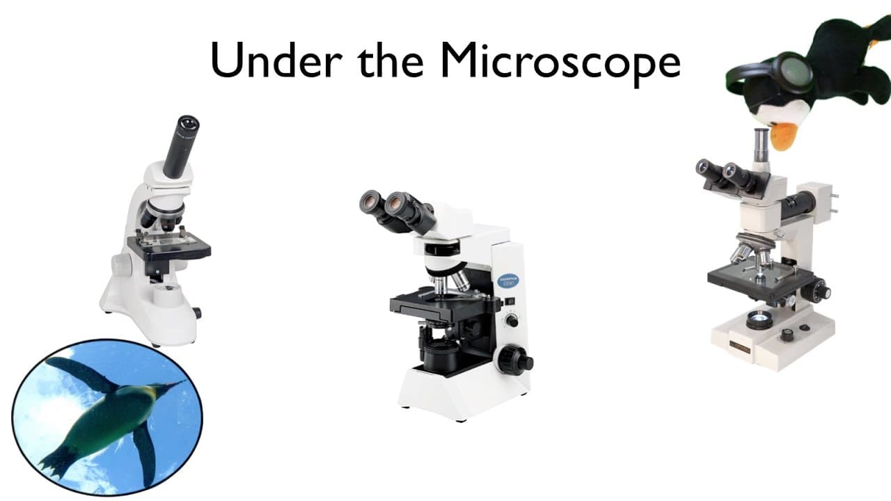
This video provides a detailed explanation of microscopes and microscopy techniques, including the parts of a compound microscope, magnification and depth of field, refraction, making a wet mount, finding objects under the microscope, and tips for protecting your eyes.
Transcript
Hello and welcome to the penguin prof channel. Today's video is part of my lab skills series. And we're going to talk about microscopes and microscopy. As always, if you find these videos helpful, I would ask that you take a moment and click those buttons below. It makes a big difference. Thanks. You want to stay tuned. We're going to be talking... Read More
Key Insights
- 🥳 Compound microscopes consist of various parts that work together to produce clear and magnified images.
- ✋ Adjusting the magnification and focus is crucial for obtaining high-quality images under the microscope.
- 💋 Wet mounts with appropriate liquid and cover slips help maintain the quality and visibility of specimens.
- 🫵 Finding objects under the microscope requires a systematic approach and understanding of field of view and magnification.
- 😃 Protecting your eyes during microscopy involves adjusting light intensity, eye relief distance, and using proper accommodation techniques.
- 🫵 Measuring the size of objects under a microscope can be done by calculating the field of view at different magnifications.
- 🥡 The inverted and 3D nature of microscope images should be taken into account while observing and interpreting specimens.
Install to Summarize YouTube Videos and Get Transcripts
Explore YouTube Video Summarizer or Get YouTube Transcript Extractor
Questions & Answers
Q: What are the main parts of a compound microscope?
The main parts of a compound microscope are the eyepiece, nosepiece, objective lenses, stage, focus knobs, and light source. Each part has a specific function in producing clear and magnified images.
Q: How do I find objects under a microscope?
Finding objects under a microscope requires systematic searching similar to a search and rescue mission. Start with a low magnification objective, scan the field of view, spot potential targets, and then increase magnification to observe more detail.
Q: How can I protect my eyes while using a microscope?
To protect your eyes, avoid excessive light, adjust the eye relief distance between your eyeball and the ocular lens, and use a near-point accommodation technique. Taking breaks and resting your eyes also helps to reduce strain.
Q: How can I measure the size of objects under a microscope without an ocular scale?
If your microscope does not have an ocular scale, you can measure the field of view under the scanning objective lens using a small clear plastic metric ruler. By calculating the magnification ratio, you can estimate the size of objects at different magnifications.
Summary & Key Takeaways
-
Microscopes are essential tools for viewing objects at a microscopic level, and the type of microscope used depends on the object being observed.
-
Compound microscopes have various parts, including the eyepiece, nosepiece, objective lenses, stage, focus knobs, and light source, all of which contribute to a clear and focused image.
-
To use a microscope, it is important to handle it carefully, adjust the magnification and focus appropriately, use wet mounts for certain specimens, and be aware of the inverted and 3D nature of microscope images.
Read in Other Languages (beta)
Share This Summary 📚
Summarize YouTube Videos and Get Video Transcripts with 1-Click
Try YouTube Summary with ChatGPT & Claude or YouTube Transcript Generator
Explore More Summaries from ThePenguinProf 📚




Summarize YouTube Videos and Get Video Transcripts with 1-Click
Try YouTube Summary with ChatGPT & Claude or YouTube Transcript Generator