Microscopy: Fluorescent Proteins (Roger Tsien)

TL;DR
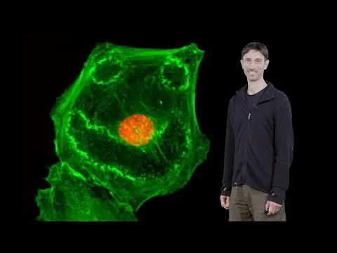
Fluorescent proteins, genetically codable and found in organisms like jellyfish and corals, have revolutionized microscopy by allowing direct visualization of molecular and cellular processes.
Transcript
Hello, I'm Roger Tsien. And I'm here today to tell you about fluorescent proteins. which have made a major impact on microscopy because they are genetically codable and provide a relatively direct link from molecular and cell biology into colors that we can directly see particularly in the light microscope but also, as you will see later on, more m... Read More
Key Insights
- 👻 Fluorescent proteins, such as GFP, have revolutionized microscopy by allowing direct visualization of molecular and cellular processes.
- 🧡 These proteins were originally discovered in jellyfish and have since been engineered and modified for a wide range of applications.
- 🧡 Fluorescent proteins from corals have expanded the available range of colors, offering new possibilities for in vivo imaging.
- ❓ Infrared fluorescent proteins provide the opportunity for imaging deeper into intact animals, enhancing our understanding of biological processes.
- ✖️ The development of multi-colored fluorescent proteins has enabled the simultaneous visualization of different proteins and cellular components within a sample.
- 🪭 Challenges in using fluorescent proteins include protein aggregation, dimerization, and proper folding, which must be carefully addressed for optimal results.
- ❓ Future advancements in fluorescent protein engineering may offer further improvements in color variety, quantum yield, and applicability in different imaging techniques.
Install to Summarize YouTube Videos and Get Transcripts
Explore YouTube Video Summarizer or Get YouTube Transcript Extractor
Questions & Answers
Q: How do fluorescent proteins work in microscopy?
Fluorescent proteins, like GFP, are genetically encoded tags that can be fused to target proteins. When exposed to specific wavelengths of light, these proteins emit a visible color, allowing researchers to visualize the location and movement of the tagged proteins within cells or tissues.
Q: What are the different colors of fluorescent proteins available?
Fluorescent proteins have been engineered to emit a range of colors, including blue, cyan, green, yellow, and even red. Each color variant is derived from specific mutations or modifications to the original protein structure.
Q: Can fluorescent proteins be used to visualize cellular processes in intact animals?
Yes, the development of infrared fluorescent proteins, which absorb and emit light at longer wavelengths, has enabled the visualization of cellular processes in intact animals. These proteins require the presence of biliverdin, a molecule produced by cells, and offer the potential for in vivo imaging with greater depth and resolution.
Q: Are there any limitations or challenges with using fluorescent proteins?
Some challenges include the potential for protein aggregation, dimerization, and improper folding. Furthermore, certain fluorescent proteins may bleach over time, requiring repeated imaging. The choice of fluorescent protein and color must be carefully considered for each specific application.
Key Insights:
- Fluorescent proteins, such as GFP, have revolutionized microscopy by allowing direct visualization of molecular and cellular processes.
- These proteins were originally discovered in jellyfish and have since been engineered and modified for a wide range of applications.
- Fluorescent proteins from corals have expanded the available range of colors, offering new possibilities for in vivo imaging.
- Infrared fluorescent proteins provide the opportunity for imaging deeper into intact animals, enhancing our understanding of biological processes.
- The development of multi-colored fluorescent proteins has enabled the simultaneous visualization of different proteins and cellular components within a sample.
- Challenges in using fluorescent proteins include protein aggregation, dimerization, and proper folding, which must be carefully addressed for optimal results.
- Future advancements in fluorescent protein engineering may offer further improvements in color variety, quantum yield, and applicability in different imaging techniques.
- The study of fluorescent proteins has not only advanced microscopy but also provides insights into biological processes, such as heme metabolism.
Summary & Key Takeaways
-
Fluorescent proteins, such as green fluorescent protein (GFP), have made a significant impact on microscopy due to their genetic codability and ability to provide visible colors.
-
These proteins were originally discovered in jellyfish and have since been engineered and optimized for various applications.
-
The discovery of fluorescent proteins from corals has expanded the range of colors available and offers potential applications in in vivo imaging of intact animals.
Read in Other Languages (beta)
Share This Summary 📚
Summarize YouTube Videos and Get Video Transcripts with 1-Click
Try YouTube Summary with ChatGPT & Claude or YouTube Transcript Generator
Explore More Summaries from iBiology Techniques 📚


Summarize YouTube Videos and Get Video Transcripts with 1-Click
Try YouTube Summary with ChatGPT & Claude or YouTube Transcript Generator