How to Create 3D Images with Confocal Microscopy

TL;DR
Confocal microscopy enables the creation of sharp 3D images by blocking out-of-focus light, unlike conventional microscopes. This is achieved using a laser to scan samples point-by-point or via a spinning disk with multiple pinholes. Each method has its strengths, with the laser scanning offering better out-of-focus rejection and the spinning disk providing faster imaging suitable for live samples.
Transcript
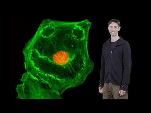
So, I am Kurt Thorn, director of the Nikon Imaging Center at UC San Francisco and I'm gonna be talking about optical sectioning and confocal microscopy and how we can use that to make 3-dimensional images of biological samples. So, our goal is to build three-dimensional images of biological samples using a microscope. And the example I'm showing he... Read More
Key Insights
- Confocal microscopy is essential for creating clear 3D images by blocking out-of-focus light.
- A laser scanning confocal microscope uses a pinhole to allow only in-focus light to reach the detector.
- Spinning disk confocal microscopy uses multiple pinholes for faster imaging, ideal for live samples.
- Laser scanning confocal microscopy provides better out-of-focus light rejection than spinning disk systems.
- Spinning disk systems are more light-efficient and suitable for low-light conditions.
- For thick samples, laser scanning is preferred due to superior out-of-focus light rejection.
- Photomultiplier tubes in confocal microscopes offer fast response times for detecting light.
- Confocal microscopy is less effective for very thick samples, where two-photon microscopy may be required.
Install to Summarize YouTube Videos and Get Transcripts
Explore YouTube Video Summarizer or Get YouTube Transcript Extractor
Questions & Answers
Q: How does confocal microscopy create 3D images?
Confocal microscopy creates 3D images by taking multiple 2D images at different focal planes and stacking them together. This is achieved by blocking out-of-focus light using a pinhole in laser scanning systems or multiple pinholes in spinning disk systems, allowing only in-focus light to be detected and reconstructed into a 3D image.
Q: What is the advantage of using a laser scanning confocal microscope?
A laser scanning confocal microscope provides excellent out-of-focus light rejection, resulting in sharper and clearer images. This is achieved by using a single pinhole to block out-of-focus light, making it ideal for imaging thick samples where precise depth information is crucial. However, it is slower than spinning disk systems due to point-by-point scanning.
Q: Why is a spinning disk confocal microscope preferred for live samples?
A spinning disk confocal microscope is preferred for live samples due to its faster imaging capabilities. It uses multiple pinholes to simultaneously illuminate and capture data from different points, allowing for rapid image acquisition. This reduces photodamage and stress on live samples, making it suitable for dynamic biological processes.
Q: How does a confocal microscope block out-of-focus light?
A confocal microscope blocks out-of-focus light by using a pinhole placed at the conjugate focal plane of the detector. In laser scanning systems, a single pinhole is used, while spinning disk systems employ multiple pinholes. This setup ensures that only light from the focal plane passes through, enhancing image clarity by eliminating blurriness from out-of-focus regions.
Q: What are the limitations of confocal microscopy?
Confocal microscopy is limited by its slower imaging speed, especially in laser scanning systems, and reduced efficiency in low-light conditions. It also struggles with very thick samples due to incomplete out-of-focus light rejection. Spinning disk systems, while faster, have limited out-of-focus rejection for thicker samples, and both systems may require alternative techniques like two-photon microscopy for extreme thicknesses.
Q: When should you use a laser scanning confocal microscope over a spinning disk system?
A laser scanning confocal microscope should be used over a spinning disk system when imaging thick samples where superior out-of-focus light rejection is necessary. It is also preferred for fixed samples where imaging speed is less critical, and precise depth resolution is required. However, it is slower and less suitable for live samples needing rapid imaging.
Q: What role do photomultiplier tubes play in confocal microscopy?
Photomultiplier tubes (PMTs) in confocal microscopy detect light with high sensitivity and speed. They convert incoming photons into electrons, amplifying the signal for accurate detection. PMTs are crucial for capturing faint fluorescence signals quickly, enabling the detailed imaging required for confocal microscopy, although they are less light-efficient compared to CCD cameras used in spinning disk systems.
Q: How does sample thickness affect the choice of confocal microscopy technique?
Sample thickness affects the choice of confocal microscopy technique by determining the need for out-of-focus light rejection and imaging speed. For thick samples, laser scanning confocal systems provide better out-of-focus rejection, while spinning disk systems are preferred for thinner samples or live specimens due to faster imaging. Extremely thick samples may require alternative methods like two-photon microscopy.
Summary & Key Takeaways
-
Confocal microscopy creates sharp 3D images by blocking out-of-focus light, unlike conventional microscopes. Laser scanning confocal microscopes use a pinhole to filter out this light, while spinning disk systems use multiple pinholes for faster imaging, suitable for live samples.
-
Laser scanning confocal microscopy offers superior out-of-focus light rejection, making it ideal for thick samples, while spinning disk systems are more light-efficient and better for low-light conditions. Each method's suitability depends on sample thickness and light conditions.
-
Photomultiplier tubes in confocal microscopes provide fast response times for detecting light, but confocal microscopy is less effective for very thick samples, where two-photon microscopy may be required. The choice between laser scanning and spinning disk depends on specific imaging needs.
Read in Other Languages (beta)
Share This Summary 📚
Summarize YouTube Videos and Get Video Transcripts with 1-Click
Try YouTube Summary with ChatGPT & Claude or YouTube Transcript Generator
Explore More Summaries from iBiology Techniques 📚


Summarize YouTube Videos and Get Video Transcripts with 1-Click
Try YouTube Summary with ChatGPT & Claude or YouTube Transcript Generator