Microscopy: Cameras and Digital Image Analysis (Nico Stuurman)

TL;DR
Explains digital cameras in microscopy and image analysis basics.
Transcript
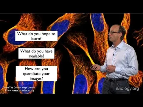
Hi I'm Nico Stuurman. I'm a staff scientist at UCSF and I prepared this lecture together with Curt Thorn, who's the director of the Nikon Imaging Center at UCSF, and I want to start out with presenting to you this nice complicated new microscope system that we have in the lab. So here we have a microscope system that we use in the lab every day. Ac... Read More
Key Insights
- Digital cameras in microscopy are essential for capturing images, which are then processed on computers for visualization and analysis. The camera's chip, containing photosensitive elements, plays a crucial role in detecting photons and converting them into digital data.
- The Nyquist-Shannon sampling theorem is critical in determining the necessary pixel size for capturing the resolution limit of a microscope. It suggests using at least two to three pixels to cover the resolution limit.
- Monochrome cameras are preferred in fluorescence microscopy due to their higher sensitivity and ability to avoid light loss, unlike color cameras that use a Bayer mask, which can reduce photon detection efficiency.
- Quantum efficiency is a key parameter in camera sensitivity, indicating the fraction of photons converted into detectable electrons. High quantum efficiency is desirable, especially in low-light conditions.
- Shot noise is an inherent noise in light signals, following a Poisson distribution. It is unavoidable, but camera-induced noise can be minimized by using advanced camera technologies and slow readout speeds.
- Digital images are matrices of numbers representing light intensity. The dynamic range, determined by bit depth, affects image quality and information content. Histograms help in adjusting image display settings for better visualization.
- File formats like TIFF are preferred for storing scientific images due to their support for lossless compression and high bit-depth data. Lossy formats like JPEG are unsuitable for scientific purposes as they can alter original data.
- Image analysis involves segmentation, where masks are created to differentiate between objects and background. This process enables quantitative measurements of image features, aiding in biological research and discoveries.
Install to Summarize YouTube Videos and Get Transcripts
Explore YouTube Video Summarizer or Get YouTube Transcript Extractor
Questions & Answers
Q: Why are monochrome cameras preferred in fluorescence microscopy?
Monochrome cameras are preferred in fluorescence microscopy because they do not use color filters that can block a significant portion of the light, unlike color cameras. This allows monochrome cameras to capture more light, which is crucial in low-light conditions typical of fluorescence microscopy. Additionally, monochrome cameras offer higher sensitivity and better signal-to-noise ratios, making them more suitable for detecting faint signals in fluorescence imaging.
Q: What is the importance of the Nyquist-Shannon sampling theorem in microscopy?
The Nyquist-Shannon sampling theorem is important in microscopy because it provides guidelines for the minimum sampling rate needed to accurately capture the resolution limit of a microscope. According to the theorem, at least two to three pixels are required to cover the smallest resolvable detail, ensuring that the digital image accurately represents the optical resolution of the microscope. This helps in selecting appropriate camera specifications for optimal image quality.
Q: How does quantum efficiency affect camera performance?
Quantum efficiency affects camera performance by determining the fraction of incoming photons that are converted into detectable electrons. A higher quantum efficiency means more photons are converted, resulting in a stronger signal and better image quality, especially in low-light conditions. Cameras with high quantum efficiency are more sensitive and can produce clearer images with less noise, which is particularly important in applications like fluorescence microscopy where light levels are low.
Q: What role do histograms play in digital image processing?
Histograms play a crucial role in digital image processing by providing a graphical representation of the distribution of pixel intensity values in an image. They help in adjusting image display settings, such as brightness and contrast, to enhance visualization. By analyzing the histogram, users can identify areas of underexposure or overexposure and adjust the intensity mapping to ensure that important details are visible, improving the interpretability of the image.
Q: Why is lossy compression not recommended for scientific images?
Lossy compression is not recommended for scientific images because it permanently removes some of the original data to reduce file size, which can alter the image content. This loss of data can compromise the accuracy and reliability of scientific analyses, as it becomes impossible to recover the original measurements. Scientific images require precise and unaltered data for accurate analysis and comparison, making lossless compression formats like TIFF more suitable for storing scientific images.
Q: What is the significance of dynamic range in digital images?
The dynamic range in digital images is significant because it determines the range of intensity values that an image can represent, affecting the image quality and information content. A higher dynamic range allows for more detailed representation of both bright and dark areas, reducing the risk of losing information in shadows or highlights. This is particularly important in scientific imaging, where capturing subtle variations in intensity can be crucial for accurate analysis and interpretation.
Q: How does segmentation aid in image analysis?
Segmentation aids in image analysis by creating masks that differentiate objects of interest from the background, allowing for quantitative measurements of specific features within an image. This process involves setting thresholds to identify regions of interest, which can then be analyzed for various parameters such as size, shape, and intensity. Segmentation is a fundamental step in image analysis, enabling researchers to extract meaningful data from complex images and facilitating automated analysis in large datasets.
Q: What are the benefits of using the TIFF file format for scientific images?
The benefits of using the TIFF file format for scientific images include its support for lossless compression, which preserves the original data without any loss, and its ability to store high bit-depth data, accommodating the wide dynamic range often required in scientific imaging. TIFF is a flexible and widely accepted format that supports both grayscale and RGB images, making it suitable for a variety of scientific applications. Its compatibility with various software tools also facilitates data sharing and analysis.
Summary & Key Takeaways
-
The lecture by Nico Stuurman covers the functioning of digital cameras in microscopy, emphasizing the importance of matching pixel size to the microscope's resolution limit using the Nyquist-Shannon theorem. Monochrome cameras are preferred for fluorescence microscopy due to their higher sensitivity compared to color cameras.
-
Quantum efficiency and shot noise are crucial factors affecting camera performance. Digital images are represented as matrices of numbers, with bit depth determining dynamic range and image quality. Histograms are useful tools for adjusting image display settings to enhance visualization.
-
TIFF is a recommended file format for storing scientific images due to its support for lossless compression and high bit-depth data. Image analysis involves segmentation to create masks for quantitative measurements, aiding in biological research and discoveries.
Read in Other Languages (beta)
Share This Summary 📚
Summarize YouTube Videos and Get Video Transcripts with 1-Click
Try YouTube Summary with ChatGPT & Claude or YouTube Transcript Generator
Explore More Summaries from iBiology Techniques 📚

Summarize YouTube Videos and Get Video Transcripts with 1-Click
Try YouTube Summary with ChatGPT & Claude or YouTube Transcript Generator