Microscopy: Dual-View Inverted Selective Plane Illumination (diSPIM) (Hari Shroff)

TL;DR
Exploring advanced microscopy techniques for studying neurodevelopment.
Transcript
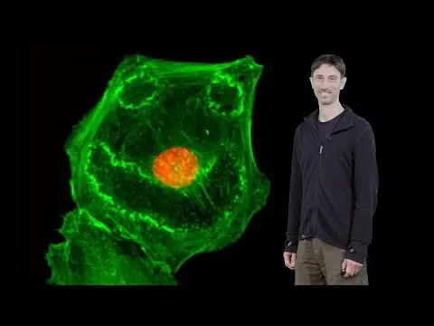
Hi. My name is Hari Shroff and I'm an investigator at the National Institute of Biomedical Imaging and Bioengineering at the NIH. My lab designs microscopes and today I'd like to tell you about a tool we developed for studying life at high spatial, temporal resolution. Before I get into the details of the microscope I'd like to describe one of the ... Read More
Key Insights
- The dual-view inverted selective plane illumination microscopy (diSPIM) offers improved spatial and temporal resolution, allowing researchers to study neurodevelopment in living organisms without causing damage.
- The C.elegans worm embryo is an ideal model for studying neurodevelopment due to its simplicity, transparency, and relatively small number of neurons and gene products.
- Traditional microscopy methods, like widefield and confocal microscopy, present challenges such as inefficiency and photodamage, limiting their use in long-term studies of embryogenesis.
- Light sheet microscopy, specifically SPIM, reduces out-of-focus light and photodamage, enabling faster and clearer imaging of embryos by illuminating only the plane of interest.
- The iSPIM system adapts SPIM for use with conventional inverted microscope bases, making it more accessible for studying samples on glass coverslips, such as C.elegans embryos.
- diSPIM enhances axial resolution by acquiring images from perpendicular directions and fusing them, achieving isotropic resolution and clearer visualization of fine structures like neuron projections.
- The diSPIM system proves effective in imaging various specimens, including GFP-EB3 labeled microtubules and GFP-labeled caveolae, with minimal photobleaching and high resolution over extended periods.
- The development and implementation of diSPIM have been a collaborative effort, with significant contributions from researchers at the NIH and beyond, highlighting the importance of teamwork in advancing microscopy technology.
Install to Summarize YouTube Videos and Get Transcripts
Explore YouTube Video Summarizer or Get YouTube Transcript Extractor
Questions & Answers
Q: What is the main purpose of the diSPIM system?
The main purpose of the diSPIM system is to provide high-resolution, non-invasive imaging of living organisms, such as the C.elegans embryo, to study neurodevelopment. It achieves this by using dual-view light sheet microscopy to reduce out-of-focus light and photodamage, allowing for extended observation of embryogenesis without harming the specimen.
Q: Why is the C.elegans worm embryo a suitable model for neurodevelopment studies?
The C.elegans worm embryo is a suitable model for neurodevelopment studies due to its simplicity, transparency, and relatively small number of neurons and gene products. It has only 302 neurons and about 20,000 gene products, making it easier to study compared to more complex organisms. Additionally, its transparency allows for the use of GFP to visualize neuronal development.
Q: How does light sheet microscopy differ from traditional microscopy methods?
Light sheet microscopy differs from traditional methods like widefield and confocal microscopy by focusing light into a single plane, reducing out-of-focus light and photodamage. This approach allows for faster imaging and clearer visualization of specimens. Unlike traditional methods, light sheet microscopy only illuminates the plane of interest, enhancing efficiency and reducing harm to the specimen.
Q: What are the advantages of using diSPIM over single-view SPIM?
diSPIM offers several advantages over single-view SPIM, including improved axial resolution and isotropic resolution by acquiring images from perpendicular directions and fusing them. This enhancement allows for clearer visualization of fine structures, such as neuron projections, and maintains high imaging speed. diSPIM also enables non-invasive imaging over extended periods with minimal photobleaching.
Q: How does the iSPIM system improve accessibility for researchers?
The iSPIM system improves accessibility for researchers by adapting SPIM for use with conventional inverted microscope bases, making it easier to study samples on glass coverslips. This design allows researchers to utilize existing equipment while benefiting from the advantages of light sheet microscopy, such as reduced photodamage and improved imaging efficiency.
Q: What challenges do traditional microscopy methods face in embryogenesis studies?
Traditional microscopy methods, like widefield and confocal microscopy, face challenges in embryogenesis studies due to inefficiency and photodamage. These methods often result in significant out-of-focus light and photobleaching, limiting their ability to continuously monitor embryos over extended periods. Consequently, they are unsuitable for long-term studies of embryogenesis.
Q: What additional specimens can be studied using the diSPIM system?
In addition to C.elegans embryos, the diSPIM system can study various specimens, such as GFP-EB3 labeled microtubules and GFP-labeled caveolae in cultured cells. The system provides high resolution and minimal photobleaching, allowing for extended observation of these specimens and enabling researchers to track small objects in three dimensions over time.
Q: Who contributed to the development and implementation of the diSPIM system?
The development and implementation of the diSPIM system have been a collaborative effort involving researchers at the NIH and beyond. Key contributors include Hari Shroff, Yicong Wu, a talented staff scientist, and Abhishek Kumar, who has been instrumental in building the microscope. This teamwork highlights the importance of collaboration in advancing microscopy technology.
Summary & Key Takeaways
-
The dual-view inverted selective plane illumination microscopy (diSPIM) is a revolutionary tool developed to study neurodevelopment with high spatial and temporal resolution. It allows for non-invasive imaging of living organisms, such as the C.elegans worm embryo, by reducing out-of-focus light and photodamage.
-
Traditional microscopy techniques like widefield and confocal microscopy have limitations that make them unsuitable for long-term embryogenesis studies. In contrast, light sheet microscopy, particularly SPIM, offers a more efficient and gentle approach by illuminating only the plane of interest.
-
The iSPIM system adapts SPIM for use with conventional inverted microscope bases, enhancing accessibility for studying samples on glass coverslips. diSPIM further improves axial resolution by acquiring images from perpendicular directions and fusing them, achieving isotropic resolution and clearer visualization of fine structures.
Read in Other Languages (beta)
Share This Summary 📚
Summarize YouTube Videos and Get Video Transcripts with 1-Click
Try YouTube Summary with ChatGPT & Claude or YouTube Transcript Generator
Explore More Summaries from iBiology Techniques 📚


Summarize YouTube Videos and Get Video Transcripts with 1-Click
Try YouTube Summary with ChatGPT & Claude or YouTube Transcript Generator