Microscopy: Differential Interference Contrast (DIC) Microscopy (Edward Salmon)
TL;DR
DIC microscopy uses polarization for high-contrast imaging.
Transcript
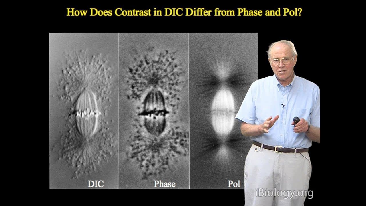
I'd like to begin this talk by asking you to look at these three images of isolated spindles, and the object here in the middle is a phase contrast image of the spindle, and as discussed in the phase contrast lecture, the contrast that we see, the dark contrast, is based on the difference in the refractive index, that being slightly greater refract... Read More
Key Insights
- DIC microscopy enhances contrast using polarizing microscopes, highlighting specimen edges rather than mass.
- The technique relies on birefringence, converting orthogonal wave retardation into visible contrast.
- DIC images use two Airy disks per point, creating directional contrast with a characteristic light-dark pattern.
- Contrast is directional, with maximum contrast along the shear direction, highlighting edges over uniform areas.
- The setup involves a dual-beam interferometer, using condenser and objective prisms to manipulate light paths.
- Compensators in DIC microscopes adjust wavefront retardation, enhancing image contrast and detail visibility.
- Nomarski prisms allow external prism placement, facilitating mechanical adjustments for compensation.
- Optimal contrast is achieved by maximizing dark edge contrast, then enhancing brightness electronically.
Install to Summarize YouTube Videos and Get Transcripts
Explore YouTube Video Summarizer or Get YouTube Transcript Extractor
Questions & Answers
Q: What is the primary advantage of DIC microscopy?
The primary advantage of DIC microscopy is its ability to produce high-contrast images that highlight the edges of specimen structures. This is achieved through the manipulation of polarized light, which enhances the visualization of fine details without the need for staining, making it particularly useful for observing live cells and transparent specimens.
Q: How does DIC microscopy differ from phase contrast microscopy?
While both DIC and phase contrast microscopy enhance contrast in transparent specimens, DIC highlights specimen edges using polarized light, creating a shadow-like effect. In contrast, phase contrast relies on differences in refractive index to produce contrast, often resulting in halos around objects. DIC provides more detailed edge definition and less halo effect.
Q: What role do Wollaston prisms play in DIC microscopy?
Wollaston prisms are crucial in DIC microscopy as they split the light beam into two orthogonally polarized components. These components are then manipulated to create interference patterns that enhance contrast. The prisms are strategically placed in the condenser and objective to ensure the beams recombine with the desired retardation, producing the characteristic DIC image.
Q: Why is understanding polarization microscopy important for DIC microscopy?
Understanding polarization microscopy is essential for DIC microscopy because DIC is essentially a variation of polarization microscopy. The technique relies on the principles of birefringence and the manipulation of polarized light to generate contrast. A foundational knowledge of how polarization affects light behavior helps in setting up and interpreting DIC images accurately.
Q: How does the shear direction affect DIC image contrast?
In DIC microscopy, the shear direction is the axis along which contrast is maximized. This direction determines how the overlapping Airy disks are oriented, affecting the appearance of light and dark edges in the image. Aligning specimen features with the shear direction enhances their visibility, making it a critical factor in obtaining clear, high-contrast images.
Q: What is the function of compensators in DIC microscopy?
Compensators in DIC microscopy are used to adjust the retardation difference between the two orthogonally polarized beams. By fine-tuning this difference, compensators enhance the contrast and brightness of the DIC image, allowing for better visualization of specimen details. They can be used in additive or subtractive modes to optimize image quality.
Q: How do Nomarski prisms improve DIC microscopy?
Nomarski prisms improve DIC microscopy by allowing the prisms to be placed outside the objective, facilitating easier adjustments. This design innovation enables the use of mechanical adjustments to control the retardation between the beams, providing flexibility in achieving the desired contrast and improving the overall usability of the DIC system.
Q: What are the challenges in setting up a DIC microscope?
Setting up a DIC microscope involves aligning multiple optical components precisely, such as the polarizer, analyzer, and DIC prisms. Achieving the correct shear and compensator settings is crucial for optimal contrast. Additionally, understanding the principles of polarization and birefringence is necessary to troubleshoot and fine-tune the system effectively for different specimens.
Summary & Key Takeaways
-
Differential Interference Contrast (DIC) microscopy is a technique that uses polarization to generate high-contrast images by highlighting specimen edges. It involves converting orthogonal wave retardation into visible contrast, using a polarizing microscope to achieve this effect.
-
The DIC setup includes a dual-beam interferometer, where condenser and objective prisms manipulate light paths to create directional contrast. This results in images characterized by a distinctive light-dark pattern, with maximum contrast along the shear direction.
-
Compensators are used to adjust wavefront retardation, enhancing image contrast and detail visibility. The Nomarski prism design allows for external prism placement, enabling mechanical adjustments. Optimal contrast is achieved by maximizing dark edge contrast and enhancing brightness electronically.
Read in Other Languages (beta)
Share This Summary 📚
Summarize YouTube Videos and Get Video Transcripts with 1-Click
Try YouTube Summary with ChatGPT & Claude or YouTube Transcript Generator
Explore More Summaries from iBiology Techniques 📚


Summarize YouTube Videos and Get Video Transcripts with 1-Click
Try YouTube Summary with ChatGPT & Claude or YouTube Transcript Generator