Microscopy: Measuring Dynamics: Fluorescent Speckle Microscopy (Clare Waterman)

TL;DR
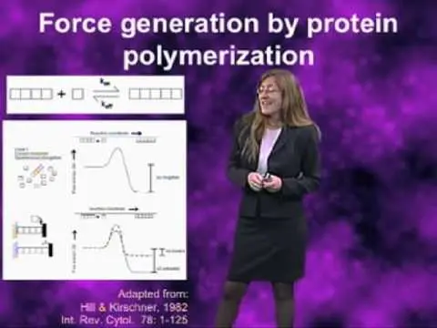
Fluorescent speckle microscopy reveals protein dynamics at high resolution.
Transcript
Hi. I'm Clare Waterman from the NIH, and I'm going to talk to you today about fluorescent speckle microscopy, which is a method that I invented by accident when I was a postdoc with Ted Salmon back in the 1990s. So, what is... light microscopy is a fabulous tool for studying the dynamics of protein macromolecular ensembles in living cells with spat... Read More
Key Insights
- Fluorescent speckle microscopy (FSM) was accidentally invented by Clare Waterman while studying microtubule dynamics.
- FSM allows researchers to study protein dynamics in living cells with high spatial and temporal precision.
- The technique overcomes the diffraction barrier of light microscopy by using low levels of fluorescently labeled proteins.
- FSM provides information on biochemical and physical properties by tracking fluorescent speckles in cells.
- The method is highly efficient, providing millions of data points per time point, necessitating computer vision tools for analysis.
- FSM can be adapted to various fluorescence microscopy techniques like confocal, spinning disk, and TIRF.
- The technique is particularly useful for studying dynamic processes such as actin treadmilling in migrating cells.
- Future developments aim to quantify absolute molecular numbers and apply FSM to new cellular contexts.
Install to Summarize YouTube Videos and Get Transcripts
Explore YouTube Video Summarizer or Get YouTube Transcript Extractor
Questions & Answers
Q: What is the main advantage of fluorescent speckle microscopy?
The main advantage of fluorescent speckle microscopy (FSM) is its ability to study protein dynamics in living cells with high spatial and temporal precision. By using low levels of fluorescently labeled proteins, FSM overcomes the diffraction barrier of light microscopy, allowing researchers to gain detailed insights into the dynamic assembly and disassembly of macromolecular structures.
Q: How was fluorescent speckle microscopy discovered?
Fluorescent speckle microscopy was discovered accidentally by Clare Waterman while she was studying microtubule dynamics in migrating epithelial cells. She noticed dim spots along microtubules instead of a continuous label, which revealed treadmilling dynamics. This observation led to the development of FSM as a technique to study protein dynamics in cells.
Q: What kind of information can FSM provide?
FSM provides detailed information on the biochemical and physical properties of proteins in living cells. By tracking fluorescent speckles, researchers can measure the rates of binding and dissociation of molecules, as well as the trajectory and velocity of materials. FSM can also probe the viscoelasticity of cellular structures, offering insights into their dynamic behavior.
Q: What are the hardware requirements for FSM?
The hardware requirements for FSM include a high-resolution microscope system capable of preventing photobleaching, efficient photon collection, and focus stability. A low noise, high dynamic range camera is essential. The microscope and detector must satisfy the Nyquist sampling criterion, and the setup should be simple, avoiding extraneous components that could affect image quality.
Q: How does FSM compare to FRAP?
FSM provides a more comprehensive view of protein dynamics compared to FRAP. While FRAP is limited to a small region and measures dynamics based on fluorescence recovery, FSM offers wide-field views and captures dynamics across the entire cell. FSM acts like conducting millions of mini-FRAP experiments simultaneously, providing detailed insights into non-steady state dynamics.
Q: What are the future directions for FSM?
Future directions for FSM include calibrating quantitative analysis software to provide absolute molecular numbers and expanding the technique's application to new cellular contexts. Researchers aim to use FSM to study processes within the nucleus, cell surface receptors, and other cytoskeletal elements. Advancements in superresolution microscopy may also enable 3-D tracking of speckle dynamics.
Q: How are speckles created in FSM?
Speckles in FSM are created by incorporating a very low fraction of fluorescently labeled proteins into a high fraction of unlabeled proteins. This random incorporation during assembly results in a speckled appearance due to differences in fluorescence intensity between adjacent regions. The technique requires bright, functional fluorescent proteins and precise control of labeling ratios.
Q: What challenges does FSM face in data analysis?
FSM generates an enormous amount of data, with millions of speckles providing information at each time point. This high data density poses a challenge for manual analysis, necessitating the use of advanced computer vision tools to efficiently extract and interpret the information. Developing robust software for quantitative analysis is crucial to fully leverage the technique's potential.
Summary & Key Takeaways
-
Fluorescent speckle microscopy (FSM) is a powerful technique that allows the study of protein dynamics in living cells with high spatial and temporal precision. By using low levels of fluorescently labeled proteins, FSM overcomes the diffraction barrier of light microscopy, providing detailed insights into macromolecular assemblies.
-
The method was accidentally discovered by Clare Waterman while studying microtubule dynamics. FSM provides biochemical and physical information by tracking fluorescent speckles, revealing processes like actin treadmilling. It generates millions of data points per time point, requiring computer vision tools for efficient data extraction.
-
FSM can be adapted to various fluorescence microscopy techniques, such as confocal, spinning disk, and TIRF. Future developments aim to quantify absolute molecular numbers and expand FSM applications to new cellular contexts, including nuclear processes and signaling pathways.
Read in Other Languages (beta)
Share This Summary 📚
Summarize YouTube Videos and Get Video Transcripts with 1-Click
Try YouTube Summary with ChatGPT & Claude or YouTube Transcript Generator
Explore More Summaries from iBiology Techniques 📚

Summarize YouTube Videos and Get Video Transcripts with 1-Click
Try YouTube Summary with ChatGPT & Claude or YouTube Transcript Generator