What Is Fluorescence Microscopy and How Does It Work?

TL;DR
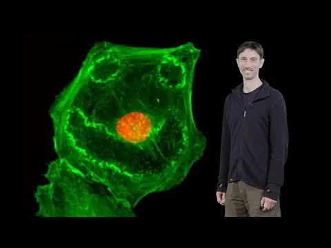
Fluorescence microscopy enhances biological imaging by using fluorescent dyes that provide high contrast and resolution, allowing for the observation of specific cellular components in living cells. This technique relies on the principles of light excitation and emission, characterized by the Stokes Shift, to deliver quantitative data on protein concentrations while facing challenges such as photobleaching.
Transcript
So much of the microscopy that we are doing now a days is fluorescence microscopy and i'll be explaining what that is in this lecture So why do we use fluorescence in the microscope? As you probably seen so far, there are two important aspects in making a great picture in your microscope One is resolution, you want high resolution, and the second t... Read More
Key Insights
- Fluorescence microscopy is crucial for high contrast and resolution in imaging, allowing specific targeting of cellular components with fluorescent dyes.
- Fluorescence can be quantitative; calibrated systems can indicate protein concentrations by fluorescence intensity.
- Fluorescence microscopy is compatible with live-cell imaging, enabling observation of dynamic processes like cell division.
- The Stokes Shift, the difference between excitation and emission wavelengths, is fundamental in fluorescence, indicating energy loss.
- Jablonski diagrams explain electron transitions in fluorescence, detailing energy absorption and emission processes.
- Quantum efficiency and brightness are key parameters of dyes, influencing their performance in fluorescence microscopy.
- Interference filters, with alternating refractive layers, are essential for distinguishing excitation from emission light in fluorescence microscopy.
- Photobleaching is a limitation of fluorescence dyes, but selecting resistant dyes and optimizing imaging conditions can mitigate its effects.
Install to Summarize YouTube Videos and Get Transcripts
Explore YouTube Video Summarizer or Get YouTube Transcript Extractor
Questions & Answers
Q: What are the main advantages of using fluorescence microscopy?
Fluorescence microscopy offers high contrast and resolution, making it possible to specifically target and visualize cellular components with fluorescent dyes. It allows for quantitative analysis by correlating fluorescence intensity with protein concentrations. Additionally, it is compatible with live-cell imaging, enabling the observation of dynamic cellular processes.
Q: How does fluorescence microscopy achieve high contrast in images?
Fluorescence microscopy achieves high contrast by using fluorescent dyes that emit light at specific wavelengths when excited by a light source. This emitted light stands out against a dark background, highlighting the labeled structures and making them easily distinguishable from the non-fluorescent surroundings, resulting in high-contrast images.
Q: What is the Stokes Shift, and why is it important in fluorescence microscopy?
The Stokes Shift is the difference between the wavelengths of absorbed and emitted light in fluorescence. It indicates an energy loss during the process, as the emitted light has a longer wavelength (lower energy) than the absorbed light. This shift is crucial for designing filters that separate excitation and emission light, ensuring clear imaging.
Q: What role do interference filters play in fluorescence microscopy?
Interference filters are essential in fluorescence microscopy for separating excitation and emission light. They consist of alternating layers with different refractive indices, allowing specific wavelengths to pass through while reflecting others. This selective filtering ensures that only the desired fluorescence is captured, improving image clarity and accuracy.
Q: What is photobleaching, and how can it be minimized in fluorescence microscopy?
Photobleaching is the irreversible loss of fluorescence in dyes due to prolonged exposure to excitation light. It can be minimized by using more stable dyes, reducing exposure time, optimizing imaging conditions, and removing oxygen from the sample environment. These strategies help preserve fluorescence and improve imaging longevity.
Q: How does fluorescence microscopy enable live-cell imaging?
Fluorescence microscopy allows live-cell imaging by using non-toxic fluorescent dyes that can label living cells without affecting their viability. This technique enables the real-time observation of cellular processes, such as cell division and protein interactions, providing valuable insights into dynamic biological activities while maintaining cell integrity.
Q: What is the significance of quantum efficiency in fluorescence microscopy?
Quantum efficiency measures the ratio of emitted photons to absorbed photons in a dye. It is a crucial parameter as it determines the brightness of the fluorescence signal. High quantum efficiency indicates that a dye efficiently converts absorbed light into fluorescence, resulting in brighter images and more accurate quantitative measurements in microscopy.
Q: What challenges are associated with using filter turrets in fluorescence microscopy?
Filter turrets in fluorescence microscopy can be slow and prone to vibrations, affecting image stability. Switching filters takes time, which can introduce delays and potential misalignment. To overcome these challenges, faster alternatives like motorized filter wheels are used, allowing rapid switching between filters and improving imaging efficiency and precision.
Summary & Key Takeaways
-
Fluorescence microscopy is a powerful technique that enhances contrast and resolution in biological imaging by using fluorescent dyes to label specific cellular components. This process allows for the observation of dynamic processes in living cells, providing quantitative data on protein concentrations.
-
Key to fluorescence microscopy are the principles of excitation and emission, characterized by the Stokes Shift. This shift represents the energy loss between absorbed and emitted light. Interference filters play a crucial role in separating excitation and emission wavelengths, ensuring clear imaging results.
-
Despite the advantages, fluorescence microscopy faces challenges like photobleaching, where dyes lose fluorescence over time. Strategies to combat this include using more stable dyes, optimizing imaging conditions, and minimizing exposure to excitation light to preserve dye fluorescence.
Read in Other Languages (beta)
Share This Summary 📚
Summarize YouTube Videos and Get Video Transcripts with 1-Click
Try YouTube Summary with ChatGPT & Claude or YouTube Transcript Generator
Explore More Summaries from iBiology Techniques 📚


Summarize YouTube Videos and Get Video Transcripts with 1-Click
Try YouTube Summary with ChatGPT & Claude or YouTube Transcript Generator